Review
. 2022 Dec 27;20(1):494.
doi: 10.1186/s12916-022-02672-y.

Obese visceral fat tissue inflammation: from protective to detrimental?

Affiliations
Review

Obese visceral fat tissue inflammation: from protective to detrimental?

Hubert Kolb. BMC Med. .

Abstract

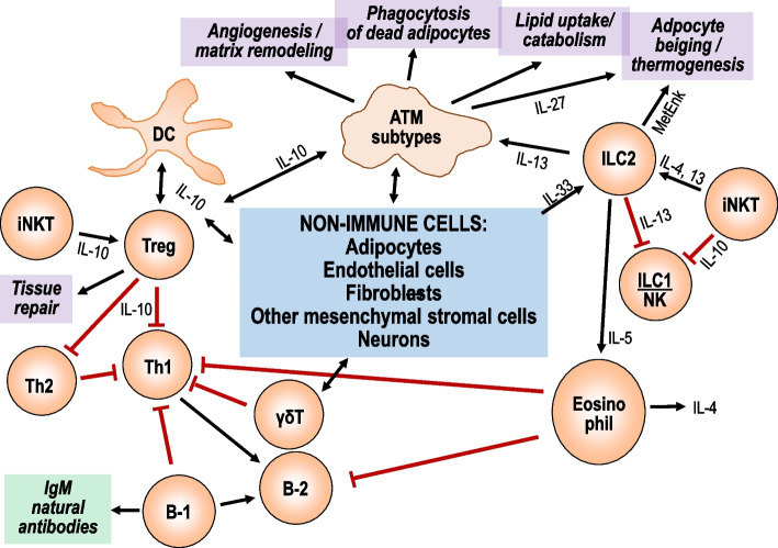
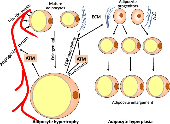
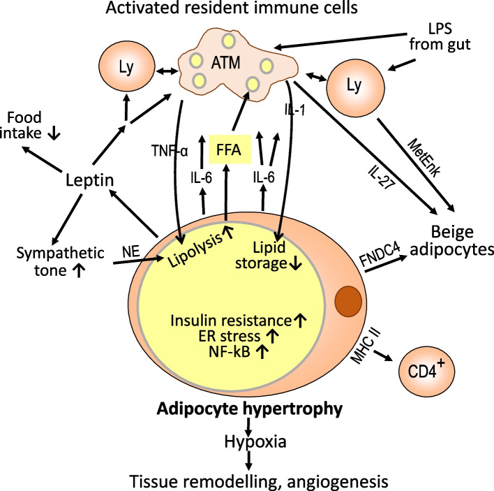
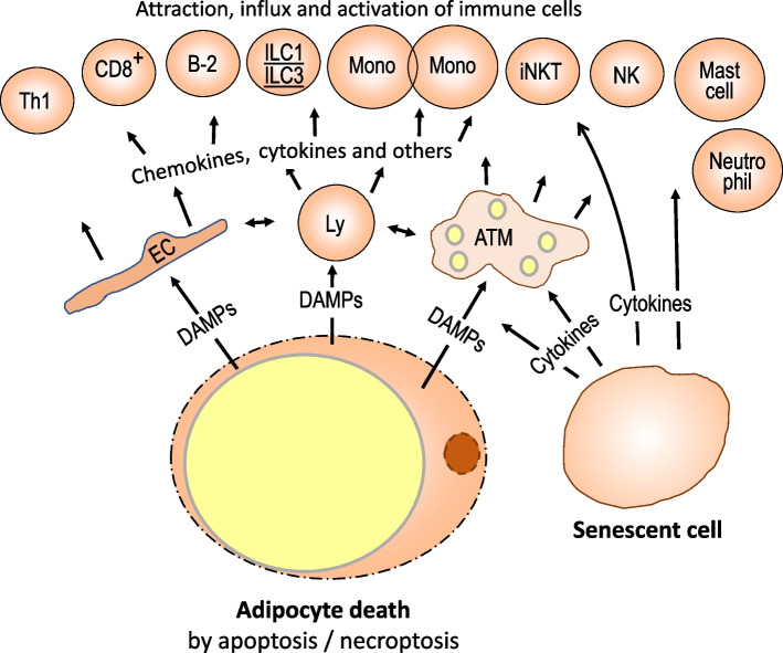
Obesity usually is accompanied by inflammation of fat tissue, with a prominent role of visceral fat. Chronic inflammation in obese fat tissue is of a lower grade than acute immune activation for clearing the tissue from an infectious agent. It is the loss of adipocyte metabolic homeostasis that causes activation of resident immune cells for supporting tissue functions and regaining homeostasis. Initially, the excess influx of lipids and glucose in the context of overnutrition is met by adipocyte growth and proliferation. Eventual lipid overload of hypertrophic adipocytes leads to endoplasmic reticulum stress and the secretion of a variety of signals causing increased sympathetic tone, lipolysis by adipocytes, lipid uptake by macrophages, matrix remodeling, angiogenesis, and immune cell activation. Pro-inflammatory signaling of adipocytes causes the resident immune system to release increased amounts of pro-inflammatory and other mediators resulting in enhanced tissue-protective responses. With chronic overnutrition, these protective actions are insufficient, and death of adipocytes as well as senescence of several tissue cell types is seen. This structural damage causes the expression or release of immunostimulatory cell components resulting in influx and activation of monocytes and many other immune cell types, with a contribution of stromal cells. Matrix remodeling and angiogenesis is further intensified as well as possibly detrimental fibrosis. The accumulation of senescent cells also may be detrimental via eventual spread of senescence state from affected to neighboring cells by the release of microRNA-containing vesicles. Obese visceral fat inflammation can be viewed as an initially protective response in order to cope with excess ambient nutrients and restore tissue homeostasis but may contribute to tissue damage at a later stage.

Keywords: Adipocyte hyperplasia; Adipocyte hypertrophy; Adipose tissue macrophages; Adiposity; Crown-like structures; Cytokines; Inflammation; Obesity; Resident immune cells; Visceral fat.


Conflict of interest statement

The author declares no competing interests.

Figures

Fig. 1
Fig. 1
Network and physiological functions of resident immune cells in lean visceral adipose tissue. In the absence of metabolic or inflammatory stress resident immune cells interact among themselves and with adipocytes and stromal cells to maintain proper tissue functions. There are no signature cytokines defining the maintenance state of resident immune cells. Rather, the concept of a buffered system applies, without polarization towards a Th1/M1- or Th2/M2-like pattern or towards another biased state of immune reactivity. Cytokines, chemokines, acute phase proteins, and other immune mediators are released in small amounts mostly from resident immune cells but also from mesenchymal stromal cells and adipocytes. Several macrophage subtypes promote matrix remodeling and angiogenesis, phagocytose dead cell and lipid aggregates, and promote adipocyte thermogenesis. ILC2 also supports adipocyte thermogenesis and stimulates physiological eosinophil functions. Regulatory T cells promote tissue repair and interact with macrophages and other immune cell types to maintain a non-inflammatory state. Low-level secretion of immune mediators by macrophages, dendritic cells, and other immune cell types such as ILC2s, iNKTs, Th2 cells, γδT cells, B-1b cells, and eosinophils helps to prevent immune cell activation. For better readability, only a few key intercellular signals are included in the scheme. ATM, adipose tissue macrophage; DC, dendritic cell; IL, interleukin; ILC, innate lymphoid cell; iNKT, innate natural killer T cell; MetEnk, methionine-enkephalin peptides; NK, natural killer cell
Fig. 2
Fig. 2
Response of visceral fat tissue to excess calories by adipocyte hypertrophy and hyperplasia. In response to high levels of circulating glucose, triglycerides, and the anabolic hormone insulin mature adipocytes take up increased amounts of nutrients and store excess energy as triglycerides in one large lipid droplet organelle. The cell size may increase 10–15-fold in diameter. Enlarged adipocytes secrete factors favoring angiogenesis and remodeling of the extracellular matrix and release of growth factors which is essential for mesenchymal stem cells, adipocyte progenitors, and preadipocytes to differentiate into lipid-storing mature adipocytes. In parallel, macrophages are stimulated to support angiogenesis and matrix remodeling. ATM, adipocyte tissue macrophages; TGs, triglycerides; Glc, glucose; ECM, extracellular matrix; Pro-inflamm., pro-inflammatory mediators
Fig. 3
Fig. 3
Local inflammation in response to disturbed adipocyte metabolic homeostasis. When enhanced lipid storage via adipocyte enlargement and differentiation of progenitor cells fails to maintain metabolic homeostasis, local inflammatory changes occur in order to dispose of excess lipid and regain metabolic control. For one, lipid-laden adipocytes experience endoplasmic reticulum stress and increased expression of NFkB leading to the release of pro-inflammatory mediators such as IL-6. Additional pro-inflammatory signals are delivered by the release of free fatty acids, leptin, lipopolysaccharides, and other products of an unbalanced microbiota in the context of a leaky gut. Activated resident immune cells release amounts of pro-inflammatory mediators sufficient to promote lipolysis and suppress lipid storage in part via induction of insulin resistance. In addition, there is an uptake of lipids by macrophages and storage in small lipid droplets. Leptin interacts with receptors in the brain to limit food intake and increase the sympathetic tone. The increased local release of noradrenaline also promotes lipolysis. Another pro-inflammatory condition results from hypoxia due to local enlargement of adipocytes. The concomitant release of enzymes and factors promoting tissue remodeling and angiogenesis may be considered a healing response. Enlarged adipocytes overexpress MHC class II antigens and appear to present antigens to CD4-positive T cells. Another pathway of limiting energy storage is the induction of adipocyte beiging by transdifferentiation or growth from progenitors and the disposal of excess energy by thermogenesis. For better readability, only a few key intercellular signals are included in the scheme. FNDC4, fibronectin type III domain-containing protein 4; FFA, free fatty acids; LPS, lipopolysaccharide; NE, norepinephrine/noradrenaline; NFkB, nuclear factor kappa B
Fig. 4
Fig. 4
Severe visceral fat tissue inflammation in response to structural disruption. Excessive enlargement of adipocytes in response to chronic overnutrition eventually causes structural damage with dying adipocytes and cell senescence as hallmarks. The phagocytotic capacity of macrophages is overwhelmed and released DAMPS strongly activate resident immune and endothelial cells resulting in the attraction of virtually all types of immune cells. Their pro-inflammatory activation also stimulates anti-inflammatory activities. Another structural change is the accumulation of senescent cells, mostly macrophages, pre-adipocytes, mature adipocytes, and endothelial cells. Senescent cells secrete pro-inflammatory mediators and enhance the accumulation of immune cells from circulation. ATM, adipose tissue macrophages; DAMP, damage-associated molecular pattern; Mono, monocytes; EC, endothelial cell; CD8+, CD8-positive T cells

References

    1. Chen L, Deng H, Cui H, Fang J, Zuo Z, Deng J, et al. Inflammatory responses and inflammation-associated diseases in organs. Oncotarget. 2018;9:7204. doi: 10.18632/oncotarget.23208. - DOI - PMC - PubMed
    1. Medzhitov R. The spectrum of inflammatory responses. Science. 2021;374:1070. doi: 10.1126/science.abi5200. - DOI - PubMed
    1. Psaila AM, Vohralik EJ, Quinlan KGR. Shades of white: new insights into tissue-resident leukocyte heterogeneity. FEBS J. 2022;289:308. doi: 10.1111/febs.15737. - DOI - PubMed
    1. Weinstock A, Brown EJ, Garabedian ML, Pena S, Sharma M, Lafaille J, et al. Single-Cell RNA Sequencing of Visceral Adipose Tissue Leukocytes Reveals that Caloric Restriction Following Obesity Promotes the Accumulation of a Distinct Macrophage Population with Features of Phagocytic Cells. Immunometabolism. 2019;1:e190008. - PMC - PubMed
    1. Schipper HS, Prakken B, Kalkhoven E, Boes M. Adipose tissue-resident immune cells: key players in immunometabolism. Trends Endocrinol Metab. 2012;23:407. doi: 10.1016/j.tem.2012.05.011. - DOI - PubMed

Publication types

LinkOut - more resources