. 2023 Apr;252(4):495-509.
doi: 10.1002/dvdy.561. Epub 2023 Jan 10.

Zebrafish model of RERE syndrome recapitulates key ophthalmic defects that are rescued by small molecule inhibitor of shh signaling

Affiliations

Zebrafish model of RERE syndrome recapitulates key ophthalmic defects that are rescued by small molecule inhibitor of shh signaling

Aman George et al. Dev Dyn. 2023 Apr.

Abstract

Background: RERE is a highly conserved transcriptional co-regulator that is associated with a human neurodevelopmental disorder with or without anomalies of the brain, eye, or heart (NEDBEH, OMIM: 616975).

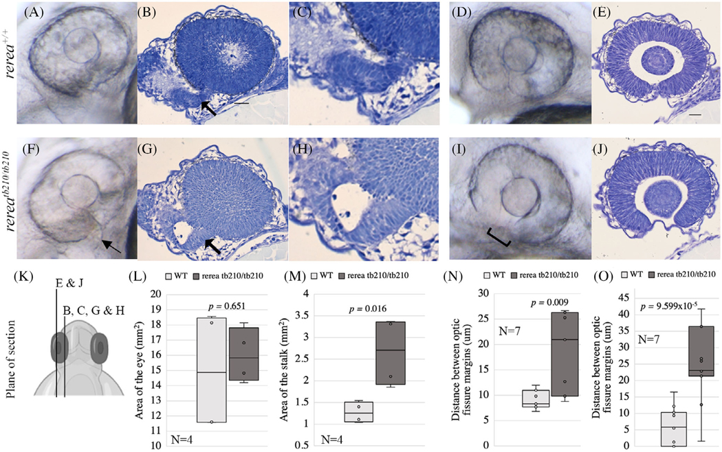
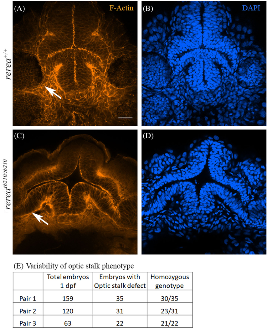
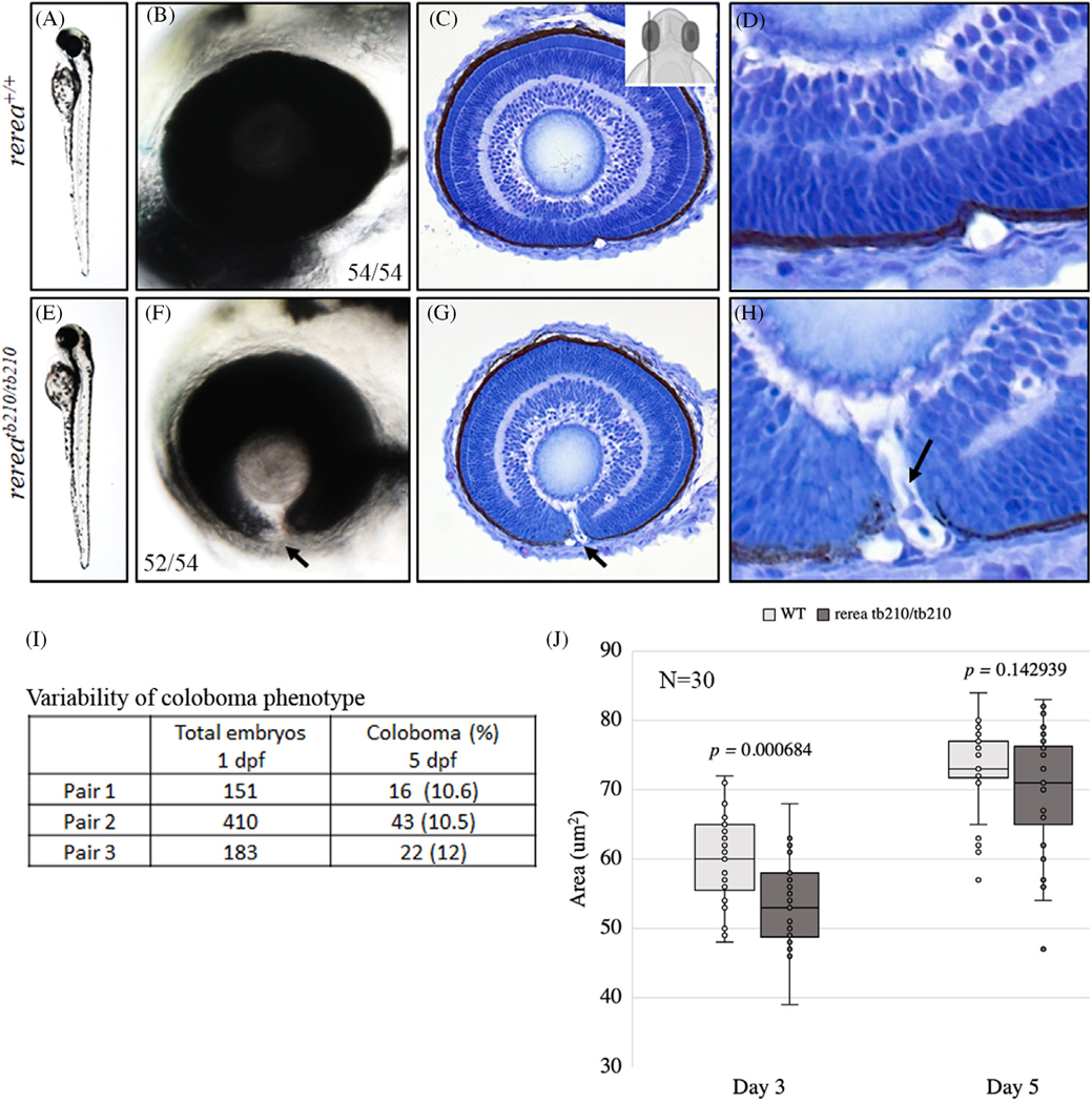
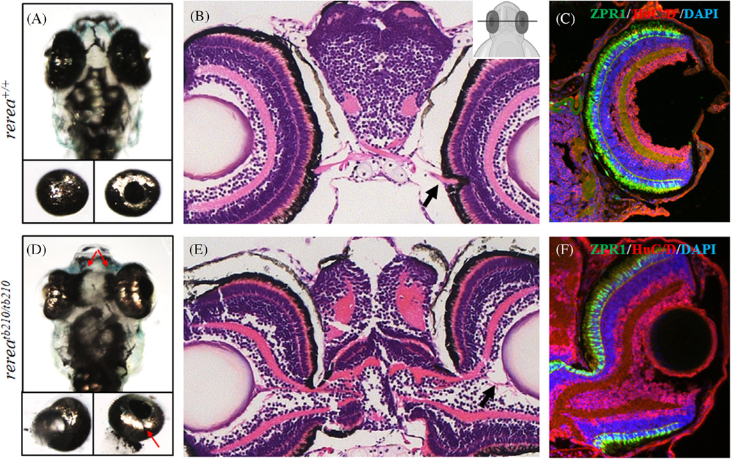
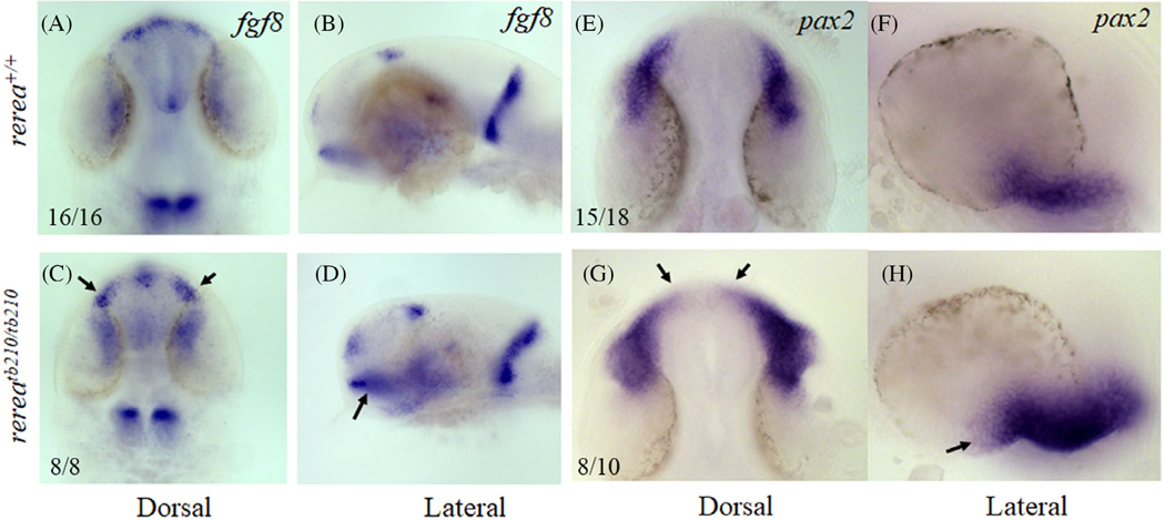
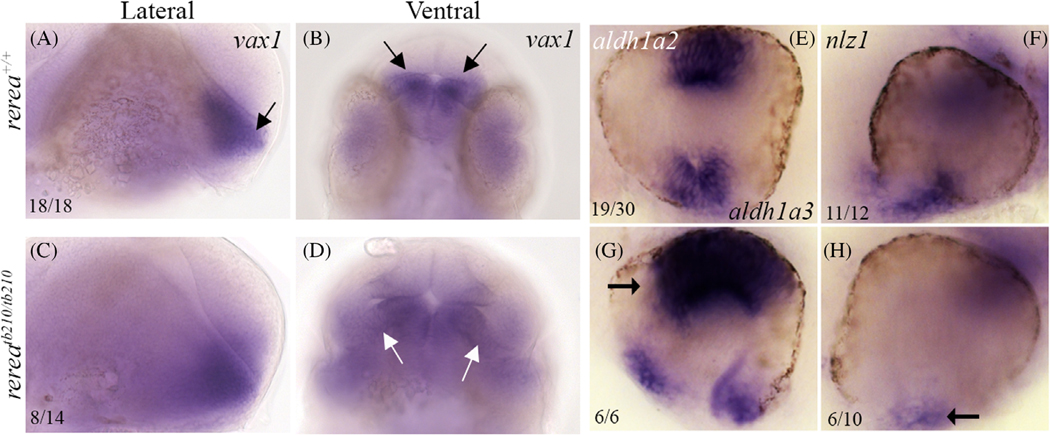
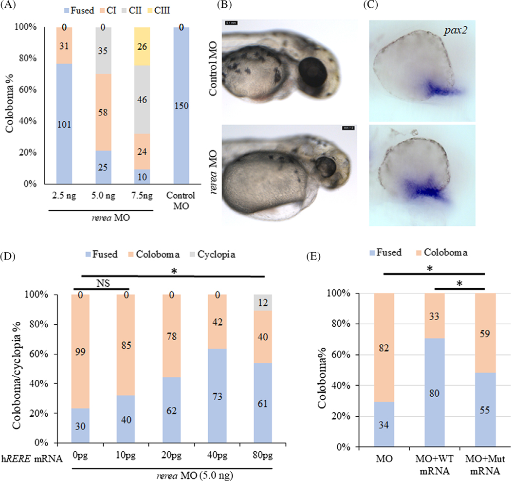
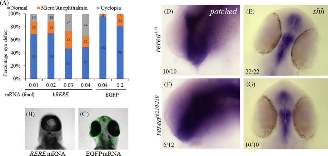
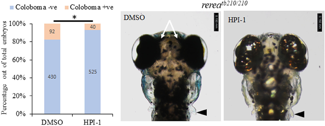
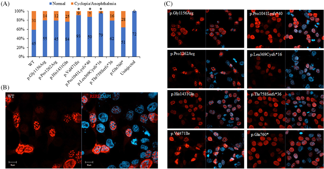
Results: We show that the zebrafish rerea mutant (babyface) robustly recapitulates optic fissure closure defects resulting from loss of RERE function, as observed in humans. These defects result from expansion of proximal retinal optic stalk (OS) and reduced expression of some of the ventral retinal fate genes due to deregulated protein signaling. Using zebrafish and cell-based assays, we determined that NEDBEH-associated human RERE variants function as hypomorphs in their ability to repress shh signaling and some exhibit abnormal nuclear localization. Inhibiting shh signaling by the protein inhibitor HPI-1 rescues coloboma, confirming our observation that coloboma in rerea mutants is indeed due to deregulation of shh signaling.

Conclusions: Zebrafish rerea mutants exhibit OS and optic fissure closure defects. The optic fissure closure defect was rescued by an shh signaling inhibitor, suggesting that this defect could arise due to deregulated shh signaling.

Keywords: RERE and SHH; coloboma; optic fissure; optic stalk.


Conflict of interest statement

CONFLICT OF INTEREST

The authors declare no competing financial interests.

Figures

FIGURE 1
FIGURE 1
Zebrafish rerea homozygous mutants exhibit enlarged optic stalk and optic fissure margins that are not apposed. Brightfield images of live fish embryos (A, F), ventro-lateral angle focused on the optic stalk [OS] and histology sections of wild-type (B, C) and rerea mutant (G, H) zebrafish embryos (24 hpf) where OS (black arrow) is present on the ventral side of the eye (lateral view). Brightfield images (D, I) and histology sections of wild-type (E) and rerea mutant (J) zebrafish embryos (24 hpf) depicting optic fissure margins that are far apart (square bracket) on the ventral side of the eye (lateral view). Schematic of plane of histology sections (K). Quantification of area of the eye (L) and optic stalk (M). Distance between optic fissure margins of brightfield images (N) and histology images (O) displayed as a box and whisker plot with dots indicating individual measurements and the horizontal bar the median value. Student’s t-test was applied for P values. Scale bar is 20 μm.
FIGURE 2
FIGURE 2
Phalloidin and DAPI stained WT (A, B) and rerea (C, D) mutant zebrafish embryos (~30 hpf) imaged from the ventral side showing optic stalk (white arrows). Table showing variability in optic stalk phenotype (E). Scale bar is 20 μm.
FIGURE 3
FIGURE 3
Zebrafish rerea homozygous mutants exhibit coloboma. Three-day post fertilization WT zebrafish embryos (A, B) exhibit fused optic fissure margins (54 genotyped embryos), whereas rerea homozygous mutants exhibit coloboma (E, F, 52/54 genotyped embryos). Sagittal histological sectioning of WT (C, D, N = 10 genotyped embryos) and rerea mutant (G, H, N = 10 genotyped embryos) zebrafish eye (3 dpf). The schematic in C gives the approximate plane of sectioning for the histologic sections. Coloboma showed variable penetrance in embryos at 5 dpf (I). Measurement of eye area (lateral view, Figure 2K) revealed significantly smaller eye at 3 dpf (P < .05) of rerea mutant larvae compared with WT. No statistical difference in eye size was observed at 5 dpf (J). Student’s t test was used to determine level of significance and P value is provided (N = 30 genotyped embryos/group).
FIGURE 4
FIGURE 4
Zebrafish rerea homozygous mutants exhibit disruption of retinal optic stalk boundary. Zebrafish larvae homozygous for rerea mutation exhibit disruption of retinal optic stalk boundary (A–C, D–F), which was further confirmed by isolation of eye cups (A, D, bottom panels) and detailed coronal histological sectioning (B, E) of 6 dpf larvae. Unfused margins of optic fissure can be observed (D bottom panel, red arrow). The presence of ectopic differentiated retinal layers (C, F) was confirmed by immunostaining with ZPR-1 (photoreceptors) and HuC/D (amacrine cells and ganglion cells).
FIGURE 5
FIGURE 5
Whole mount in situ hybridization staining of zebrafish rerea homozygous mutants exhibit expanded expression domains of optic stalk markers fgf8 and pax2a. Dorsal and lateral views of 24 hpf zebrafish embryos stained with fgf8 (A–D) and pax2a (E–H) exhibited expanded expression domains (arrows) in rerea mutants (C, D, G, H) as compared with WT (A, B, E, F) embryos
FIGURE 6
FIGURE 6
Zebrafish rerea mutants exhibit disruption of proximo-distal patterning. Whole mount in situ hybridization staining of 24 hpf zebrafish embryos with vax1, aldh1a2, aldh1a3, and nlz1 probes. Numbers of embryos with patterns of staining similar to that pictured are given in each panel
FIGURE 7
FIGURE 7
Morpholino-mediated knockdown of rerea causes coloboma. Increasing concentrations of rerea morpholino was used for knocking down rerea expression (A). Severity of coloboma is denoted by: CI-mild, CII-moderate, CIII-severe. Zebrafish embryos injected with rerea morpholino consistently exhibited coloboma at 48 hpf compared with embryos injected with control morpholino (B). Representative whole mount in situ hybridization staining with pax2a in control and rerea morpholino injected embryos 24 hpf (n ~20 embryos each) (C). WT human RERE mRNA rescues coloboma in rerea morphant fish embryos (D), this rescue was observed to be statistically significant (P < .05) for 20, 40, and 80 pg of mRNA injected, but not for 10 pg. Equal amounts of the human RERE WT and mutant (c.3466 G > A, p.Gly1156Arg) mRNA (40 pg) can rescue coloboma in rerea morphants (P < .05), but the mutant mRNA is not as efficient as WT mRNA (P < .05, E). Student’s t test was used to determine level of significance and “*” denotes significant difference. NS, not significant
FIGURE 8
FIGURE 8
Injection of WT human RERE mRNA results in cyclopia. Injecting increasing concentration of human RERE mRNA results in dose-dependent increase in cyclopia phenotype (A–C). Whole mount in situ hybridization staining of 24 hpf WT and rerea mutant zebrafish embryos with patched2 (D, F) and shha (E, G) probes. Representative images with numbers of embryos showing this pattern are given in each panel.
FIGURE 9
FIGURE 9
Shh signaling inhibitor HPI-1 rescues the blowout coloboma phenotype in rerea mutant zebrafish embryos. Treatment of rerea mutant zebrafish embryos with HPI-1 (5 μM) reduces the occurrence of coloboma phenotype from 71.3% to 26.3% (Student’s t test, P < .05) coloboma in homozygous mutant embryos.
FIGURE 10
FIGURE 10
Human RERE variants exhibit hypomorphic activity in vivo and mis-localized expression in vitro. In vivo assay to determine the efficiency of human RERE variants (80 pg) to cause cyclopia in zebrafish model (A, χ2 test was used to determine level of significance and “*” denotes significant difference). In vitro assay to determine the nuclear localization pattern of human RERE wild type (WT, B) and mutant proteins (C) in HEK293 cells. Scale bar is 10 μm.

Similar articles

References

    1. Fregeau B, Kim BJ, Hernández-García A, et al. De novo mutations of RERE cause a genetic syndrome with features that overlap those associated with proximal 1p36 deletions. Am J Hum Genet. 2016;98(5):963–970. doi:10.1016/j.ajhg.2016.03.002 - DOI - PMC - PubMed
    1. Asai Y, Chan DK, Starr CJ, Kappler JA, Kollmar R, Hudspeth AJ. Mutation of the atrophin2 gene in the zebrafish disrupts signaling by fibroblast growth factor during development of the inner ear. Proc Natl Acad Sci. 2006;103(24):9069–9074. doi:10.1073/pnas.0603453103 - DOI - PMC - PubMed
    1. Plaster N, Sonntag C, Schilling TF, Hammerschmidt M. REREa/Atrophin-2 interacts with histone deacetylase and Fgf8 signaling to regulate multiple processes of zebrafish development. Dev Dyn. 2007;236(7):1891–1904. doi:10.1002/dvdy.21196 - DOI - PubMed
    1. Neuhauss SC, Biehlmaier O, Seeliger MW, et al. Genetic disorders of vision revealed by a behavioral screen of 400 essential loci in zebrafish. J Neurosci. 1999;19(19):8603–8615. - PMC - PubMed
    1. Wang L, Charroux B, Kerridge S, Tsai CC. Atrophin recruits HDAC1/2 and G9a to modify histone H3K9 and to determine cell fates. EMBO Rep. 2008;9(6):555–562. doi:10.1038/embor.2008.67 - DOI - PMC - PubMed

Publication types

LinkOut - more resources