

Clinical characteristics of patients with epiretinal membrane-Foveoschisis
- PMID: 36576570
- PMCID: PMC10198886
- DOI: 10.1007/s00417-022-05940-y
Clinical characteristics of patients with epiretinal membrane-Foveoschisis
Abstract
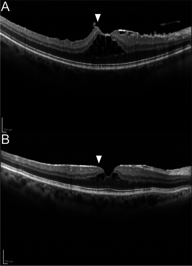
Purpose: The purpose of this study is to investigate the clinical and morphological characteristics of epiretinal membrane (ERM)-Foveoschisis.
Methods: Medical charts of 2088 patients diagnosed with idiopathic ERM were screened and eyes with ERM-Foveoschisis were included. All eyes underwent a complete ophthalmological examination including spectral domain optical coherence tomography (SD-OCT). OCT features and best corrected visual acuity (BCVA) were analysed. ERM-Foveoschisis was defined as open, closed, elevated or flat based on the OCT features. Ellipsoidal zone (EZ) abnormality, intraretinal cystoid spaces, central foveal thickness (CFT), posterior vitreous detachment (PVD) and lens status were assessed.
Results: One hundred-sixty-six patients (175 eyes) (72% female, mean age 70.46 years) were included. Incidence of ERM-Foveoschisis was 6.7%. Open type was seen in 86.8% and had a significantly better mean BCVA than closed type (p = 0.01). No statistically significant difference of mean BCVA was noted between the elevated and flat types. Mean BCVA was significantly lower in eyes with EZ abnormality (p = 0.03) and eyes with intraretinal cystoid spaces (p = 0.02). Patients with 'closed' ERM-Foveoschisis showed a significant higher median CFT than 'open' ERM-Foveoschisis (respectively, 364 µm and 176 µm, p < 0.001). A total of 81.9% eyes had PVD.
Conclusion: We differentiated four morphological types of ERM-Foveoschisis based on the OCT examination. Closed ERM-Foveoschisis presented with a higher CFT and lower BCVA than the open type. ERM-Foveoschisis with cystoid intraretinal spaces presented with a lower BCVA. The impact of the morphological types of the ERM-Foveoschisis on the clinical course and for therapy decision requires further long-term studies.
Keywords: Degenerative lamellar macular hole; Epiretinal membrane; Foveoschisis; Lamellar macular hole; Optical coherence tomography; Tractional lamellar hole.
© 2022. The Author(s).
Conflict of interest statement
The authors declare no competing interests.
Figures

References
-
- Meuer SM, Myers CE, Klein BE, Swift MK, Huang Y, Gangaputra S, Pak JW, Danis RP, Klein R. The epidemiology of vitreoretinal interface abnormalities as detected by spectral-domain optical coherence tomography: the beaver dam eye study. Ophthalmology. 2015;122:787–795. doi: 10.1016/j.ophtha.2014.10.014. - DOI - PMC - PubMed
MeSH terms
LinkOut - more resources
Full Text Sources
