

IgA-dominant glomerulonephritis with DNAJB9-negative fibrillar polytypic immunoglobulin deposits in the subepithelium
- PMID: 36576710
- PMCID: PMC10393911
- DOI: 10.1007/s13730-022-00759-2
IgA-dominant glomerulonephritis with DNAJB9-negative fibrillar polytypic immunoglobulin deposits in the subepithelium
Abstract
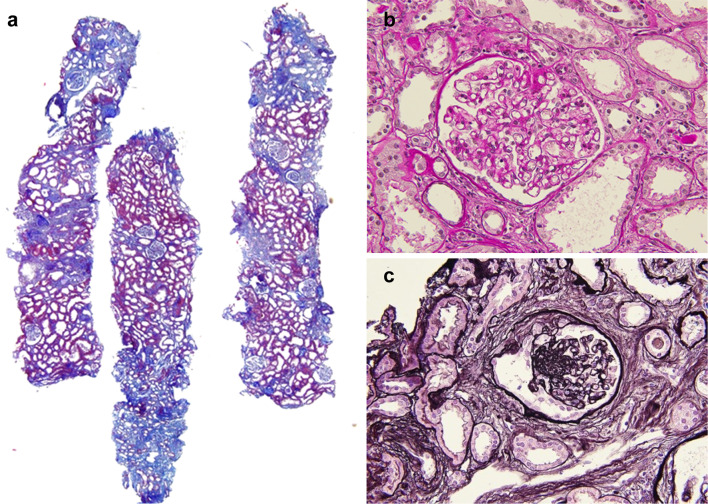
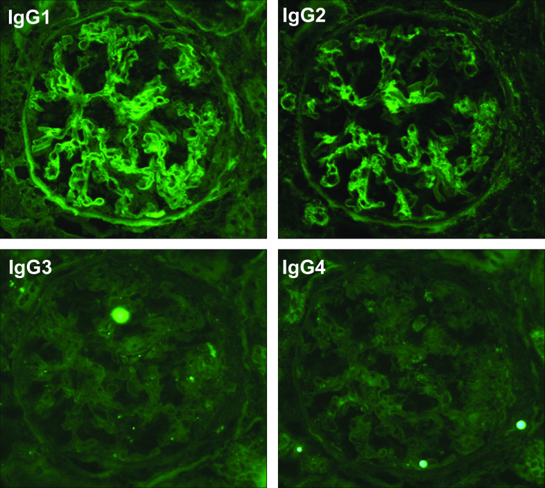
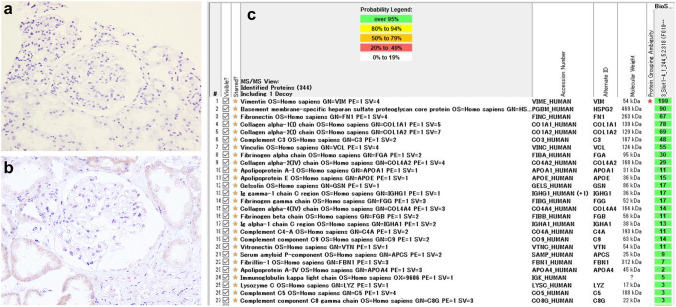
Fibrillary glomerulonephritis (FGN), a rare disease is pathologically characterized by glomerular fibril accumulation ranging from 12 to 24 nm in diameter with negative Congo red staining. Recently, the identification of DnaJ homolog subfamily B member 9 (DNAJB9) as a highly sensitive and specific marker for FGN has revolutionized diagnosis of this disease. However, few recent studies have reported DNAJB9-negative glomerulonephritis with fibrillar deposits. As such, it remains unclear whether DNAJB9-negative cases can be considered equivalent to FGN. Here, we report the case of a 70-year-old woman who developed renal impairment and nephrotic-range proteinuria. Renal biopsy and pathological examination revealed focal glomerulonephritis with fibrocellular crescents. Immunofluorescence microscopy showed IgA-dominant deposition of polytypic IgG in the glomerulus. Electron microscopy revealed hump-like subepithelial electron dense deposits with fibrils of 15-25 nm in diameter. These findings were consistent with FGN; thus, Congo red and direct fast scarlet (DFS) staining, and immunohistochemistry for DNAJB9 were performed. In addition to negative Congo red/DFS/DNAJB9 staining, laser microdissection (LMD) and liquid chromatography-tandem mass spectrometry (LC-MS/MS) resulted negative for DNAJB9, which is a highly sensitive and specific marker for FGN. The patient's renal function further declined, prompting administration of rituximab weekly for 2 weeks, similar to the treatment for FGN. This is a unique case of IgA-dominant glomerulonephritis with DNAJB9-negative fibrillar polytypic immunoglobulin deposits in the subepithelium, unlike previous DNAJB9-negative cases. Thus, DNAJB9-negative cases diagnosed based on accurate electron microscopic evaluation must be gathered, and LMD and LC-MS/MS must be used to analyze the organized fibrillar deposits to reveal the disease entity.
Keywords: DNAJB9; Fibrillar glomerulonephritis; IgA-dominant deposits; Kidney biopsy; Mass spectrometry; Subepithelial deposits.
© 2022. The Author(s) under exclusive licence to The Japan Society of Nephrology.
Conflict of interest statement
All the authors have declared no competing interest.
Figures


References
Publication types
MeSH terms
Substances
LinkOut - more resources
Full Text Sources
Research Materials
Miscellaneous
