. 2022 Dec 28;24(1):282.
doi: 10.1186/s13075-022-02971-y.

Early removal of the infrapatellar fat pad/synovium complex beneficially alters the pathogenesis of moderate stage idiopathic knee osteoarthritis in male Dunkin Hartley guinea pigs

Affiliations

Early removal of the infrapatellar fat pad/synovium complex beneficially alters the pathogenesis of moderate stage idiopathic knee osteoarthritis in male Dunkin Hartley guinea pigs

Maryam F Afzali et al. Arthritis Res Ther. .

Abstract

Background: The infrapatellar fat pad (IFP) is the largest adipose deposit in the knee; however, its contributions to the homeostasis of this organ remain undefined. To determine the influence of the IFP and its associated synovium (IFP/synovium complex or IFP/SC) on joint health, this study evaluated the progression of osteoarthritis (OA) following excision of this unit in a rodent model of naturally-occurring disease.

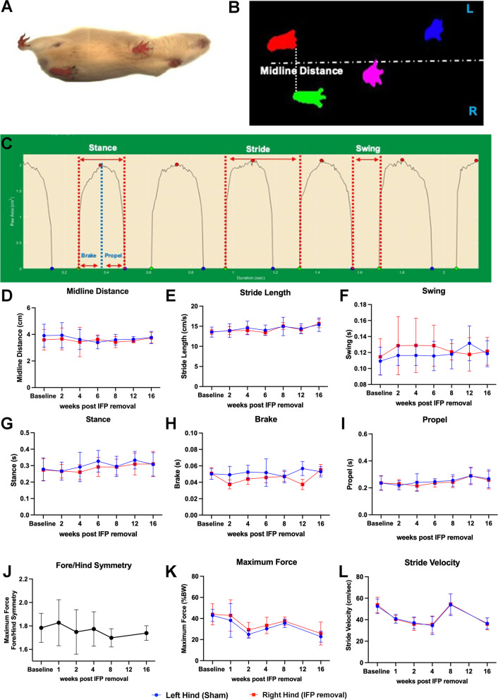
Methods: Male Dunkin-Hartley guinea pigs (n=18) received surgical removal of the IFP in one knee at 3 months of age; contralateral knees received sham surgery as matched internal controls. Mobility and gait assessments were performed prior to IFP/SC removal and monthly thereafter. Animals were harvested at 7 months of age. Ten set of these knees were processed for microcomputed tomography (microCT), histopathology, transcript expression analyses, and immunohistochemistry (IHC); 8 sets of knees were dedicated to microCT and biomechanical testing (material properties of knee joints tissues and anterior drawer laxity).

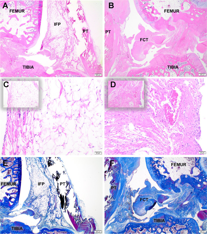
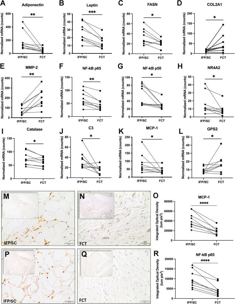
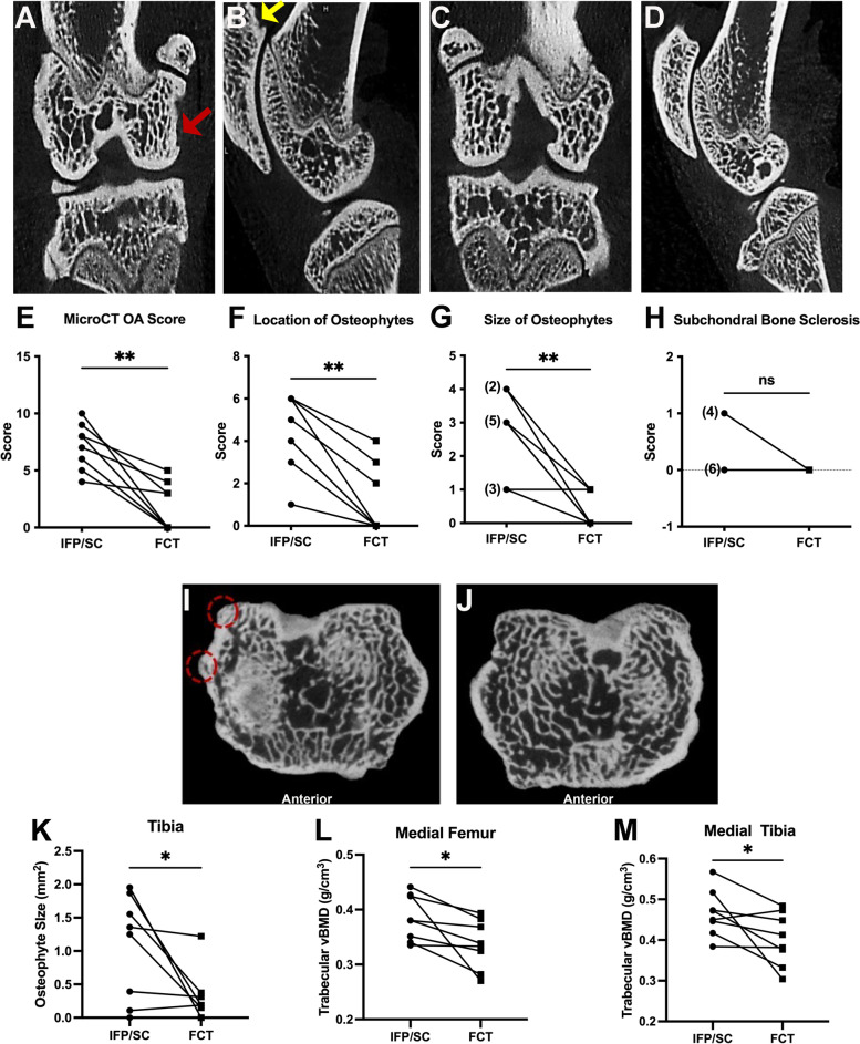
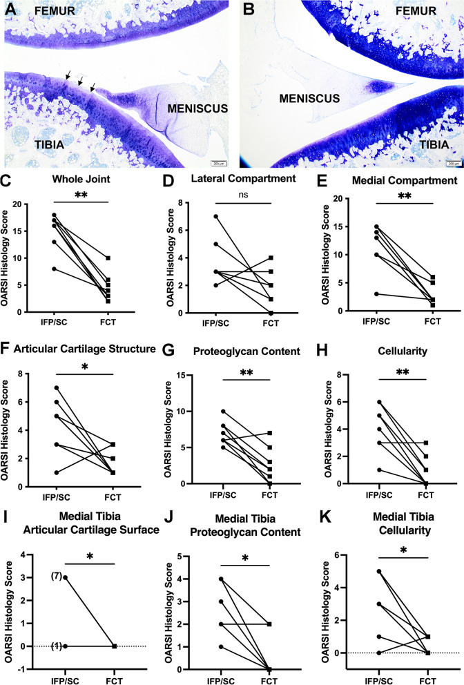
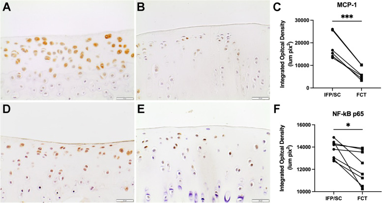
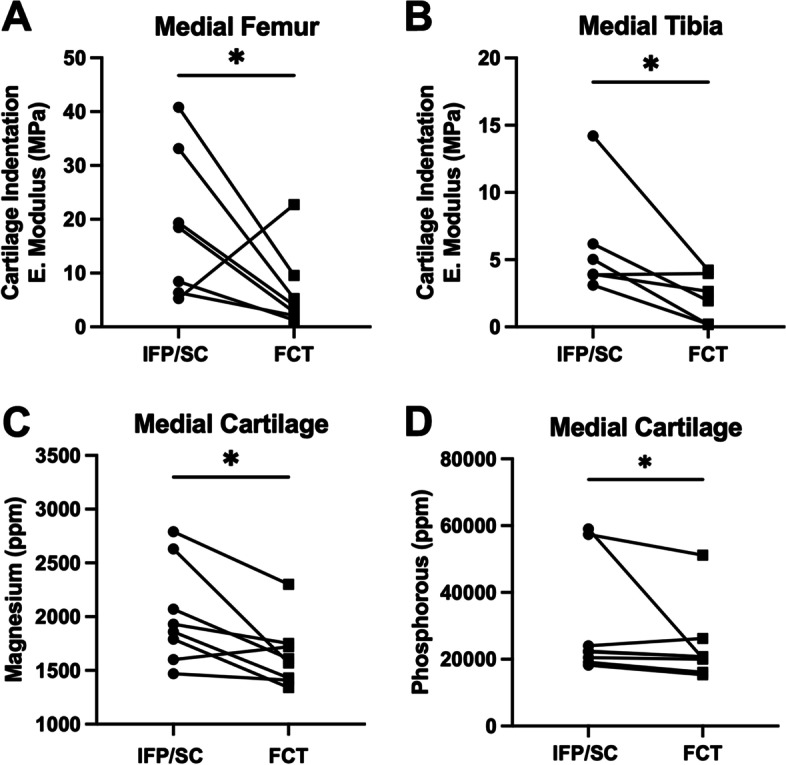
Results: Fibrous connective tissue (FCT) developed in place of the native adipose depot. Gait demonstrated no significant differences between IFP/SC removal and contralateral hindlimbs. MicroCT OA scores were improved in knees containing the FCT. Quantitatively, IFP/SC-containing knees had more osteophyte development and increased trabecular volume bone mineral density (vBMD) in femora and tibiae. Histopathology confirmed maintenance of articular cartilage structure, proteoglycan content, and chondrocyte cellularity in FCT-containing knees. Transcript analyses revealed decreased expression of adipose-related molecules and select inflammatory mediators in FCTs compared to IFP/SCs. This was verified via IHC for two key inflammatory agents. The medial articular cartilage in knees with native IFP/SCs showed an increase in equilibrium modulus, which correlated with increased amounts of magnesium and phosphorus.

Discussion/conclusion: Formation of the FCT resulted in reduced OA-associated changes in both bone and cartilage. This benefit may be associated with: a decrease in inflammatory mediators at transcript and protein levels; and/or improved biomechanical properties. Thus, the IFP/SC may play a role in the pathogenesis of knee OA in this strain, with removal prior to disease onset appearing to have short-term benefits.

Keywords: Biomechanics; Gait; Hartley guinea pig; Inflammation; Infrapatellar fat pad/synovium complex; Osteoarthritis; Trace elements.


Conflict of interest statement

No authors have any conflicts of interest to disclose for this work.

Figures

Fig. 1
Fig. 1
Mobility assessments. (A) Schematic of treadmill-based gait analysis measurements. (B) Video still image showing a guinea pig running on a transparent treadmill (viewed from below) at 45 cm/s. Placement for each paw is detected from the video illustrated in A. (C) Paw area in contact with treadmill surface over time for a representative single paw (left hind limb), from which multiple stride indices can be obtained (labeled). Longitudinal data of IFP (blue) and replacement tissue (red) contralateral limb measurements of midline distance (D), stride length (E), swing (F), stance (G), brake (H), and propel time (I). Voluntary weight-bearing assessments between fore/hind symmetry (J), maximum force (K), and stride velocity (L). P-values represent the significance of two-way ANOVA (or mixed model) with Tukey’s post hoc test analyses
Fig. 2
Fig. 2
Characterization of IFP/SC and replacement tissue. Representative mid-sagittal photomicrographs of a stifle joint from a control (A, E) and IFP/SC removal (B, F) guinea pig; H&E (A, B), and Masson’s trichrome (E, F), 2× objective. (A, C) Control knee joint from a 7-month-old guinea pig depicting the normal histoanatomic location of the IFP/SC in its native state. (B, D) Knee joint from 7-month-old guinea pig four months after IFP/SC removal. The IFP/SC (C) is replaced with dense fibrous connective tissue (FCT) 20× objective for the main photo; 10× objective for inset (D)
Fig. 3
Fig. 3
Transcript and protein expression analyses. Normalized mRNA counts for adiponectin (A), leptin (B), FASN (C), COL2A1 (D), MMP-2 (E), NF-kB p65 (F), NF-kBp50 (G), NR4A2 (H), catalase (I), C3 (J), MCP-1 (K), and GPS2 (L) in IFP/SC and FCT of knee joints. Immunohistochemistry for MCP-1 expression in IFP/SC  (M; left hind limb) and FCT (N; right hind limb). MCP-1 is a cytokine regulated by the NF-kBp65 pathway; expression in the FCT was reduced compared to the native IFP/SC. (O) Figure representing quantitative analysis of MCP-1-stained tissue subtracted from IgG control tissue for all samples. NF-kB is a transcription factor that is known for regulating inflammatory responses within inflammatory cells; protein expression is increased in IFP/SC (P; left hind limb) versus FCT (Q; right hind limb). (R) Figure representing quantitative analysis of p65 stained tissue subtracted from IgG control tissue for all samples. P-values were determined by paired t-test for non-parametric Wilcoxon test× for non-normally distributed data. ***P <0.0005, **P<0.005, and *P<0.05
Fig. 4
Fig. 4
Clinical and quantitative MicroCT. Representative photos from microCT evaluation of knee joints using the published scoring system. (A) Coronal and sagittal (B) sections of an IFP/SC-containing knee. Subchondral bone sclerosis is present on the medial femoral condyle (coronal section, red arrow). An osteophyte is highlighted on the proximal patella (sagittal section, yellow arrow). (C) Coronal and sagittal sections of the FCT limb, which is relatively unaffected. (E) The clinical microCT OA score demonstrated a significant decrease in bony changes in the FCT knees (P= 0.0020). Contributions to whole joint microCT OA score components included location of osteophytes (F), size of osteophytes (G), and subchondral bone sclerosis (H). Cross-section of tibiae containing either IFP/SC (I) or FCT (J); osteophytes are highlighted on the medial posterior tibia (red dashed circles). (K) Osteophyte volume mm3 was significantly decreased in FCT versus IFP/SC knees. Trabecular vBMD (g/cm3) of the medial femora (L) and tibiae (M) were decreased by the FCT when compared to the IFP/SC. Normally distributed data with similar variance were compared using parametric t test. Data with non-Gaussian distribution were compared using non-parametric Wilcoxon× matched – pairs signed rank test. **P<0.005 and *P<0.05
Fig. 5
Fig. 5
OARSI Score. Representative photomicrographs of toluidine blue-stained sections from medial compartments for OARSI scoring. (A) Irregular articular surface with mild fibrillation and proteoglycan loss is present in the superficial zone of the tibia from the knee containing the native IFP/SC. (B) The contralateral knee from the same animal exhibits a smooth cartilage surface and very mild proteoglycan loss. (C) OARSI whole joint OA score confirmed a significant statistical difference in cartilage and proteoglycan change. Contributions to whole joint OARSI score components included the lateral compartment (D) OARSI score, which was not significantly different, and medial compartment (E) mean OARSI histologic score, which was significantly different. Contributions to the medial compartment included articular cartilage structure (F), proteoglycan content (G), and cellularity (H). More specifically, medial tibia OARSI score of articular cartilage surface (I), proteoglycan content (J), and cellularity (K) contributed to the significant decrease in histolopathology by the FCT when compared to the IFP/SC. Normally distributed data with similar variance were compared using parametric t test. Data with non-Gaussian distribution were compared using non-parametric Wilcoxon× matched – pairs signed rank test. **P<0.005 and *P<0.05
Fig. 6
Fig. 6
MCP-1 and NF-kB p65 immunohistochemistry of medial tibia plateau (MTP) articular cartilage. 40× objective representative images of MTP articular cartilage immunostained for MCP-1 from IFP/SC (A) and FCT-containing knees (B) of the same animal. (C) Figure representing quantitative analysis of MCP-1-stained tissue subtracted from IgG control tissue for all samples. IHC for NF-kB p65 was also significantly decreased in FCT knees compared (E) to IFP/SC knees (D). (G) Figure representing quantitative analysis of p65 stained tissue subtracted from IgG control tissue for all samples. Data was normally distributed and compared using parametric ratio paired t test. *** P < 0.0005 and * P< 0.05
Fig. 7
Fig. 7
Indentation and trace element concentration of medial cartilage. Equilibrium elastic modulus of the medial femur (A) and medial tibia (B) were lower in FCT-containing knees when compared to the IFP/SC. Medial cartilage analyzed for trace element revealed a lower concentration of Mg (C) and P (D) in cartilage from FCT knees compared to cartilage collected from IFP/SC knees. Data was normally distributed and compared using parametric ratio paired t test. * P< 0.05

Similar articles

Cited by

References

    1. March L, Smith EU, Hoy DG, Cross MJ, Sanchez-Riera L, Blyth F, Buchbinder R, Vos T, Woolf AD. Burden of disability due to musculoskeletal (MSK) disorders. Best Pract Res Clin Rheumatol. 2014;28(3):353–366. doi: 10.1016/j.berh.2014.08.002. - DOI - PubMed
    1. Sowers MR, Karvonen-Gutierrez CA. The evolving role of obesity in knee osteoarthritis. Curr Opin Rheumatol. 2010;22(5):533–537. doi: 10.1097/BOR.0b013e32833b4682. - DOI - PMC - PubMed
    1. Katz JN, Brownlee SA, Jones MH. The role of arthroscopy in the management of knee osteoarthritis. Best Pract Res Clin Rheumatol. 2014;28(1):143–156. doi: 10.1016/j.berh.2014.01.008. - DOI - PMC - PubMed
    1. Kotlarz H, Gunnarsson CL, Fang H, Rizzo JA. Insurer and out-of-pocket costs of osteoarthritis in the US: evidence from national survey data. Arthritis Rheum. 2009;60(12):3546–3553. doi: 10.1002/art.24984. - DOI - PubMed
    1. Trippel SB, Ghivizzani SC, Nixon AJ. Gene-based approaches for the repair of articular cartilage. Gene Ther. 2004;11(4):351–359. doi: 10.1038/sj.gt.3302201. - DOI - PubMed

Publication types

Substances

LinkOut - more resources