

Influence of the contact area of the sub-antral space with sinus bone and the Schneiderian membrane on osteogenesis in lateral window sinus elevation surgery: a prospective experiment
- PMID: 36578061
- PMCID: PMC9798614
- DOI: 10.1186/s12903-022-02694-1
Influence of the contact area of the sub-antral space with sinus bone and the Schneiderian membrane on osteogenesis in lateral window sinus elevation surgery: a prospective experiment
Abstract
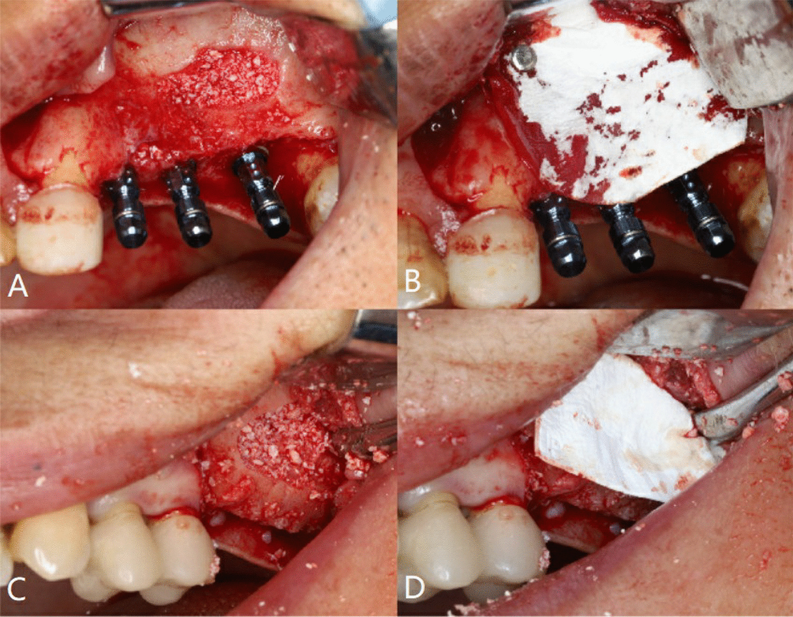
Background: Osteogenesis of lateral window sinus elevation surgery is the key to placement of the subsequent implant, excessive collapse of the sub-antral space may adversely affect long-term stability of implants. At present, few studies focus on the influence of the contact area of the sub-antral space on osteogenesis. This study evaluated whether the change in the contact area of the sub-antral space with maxillary sinus bone and the Schneiderian membrane can affect osteogenesis.
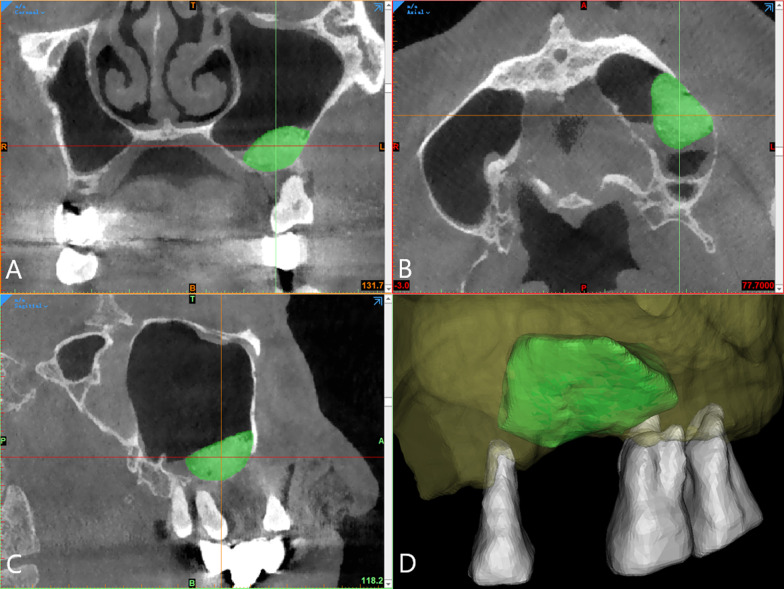
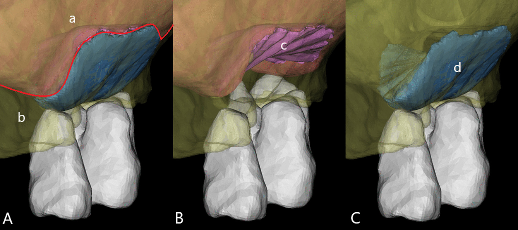
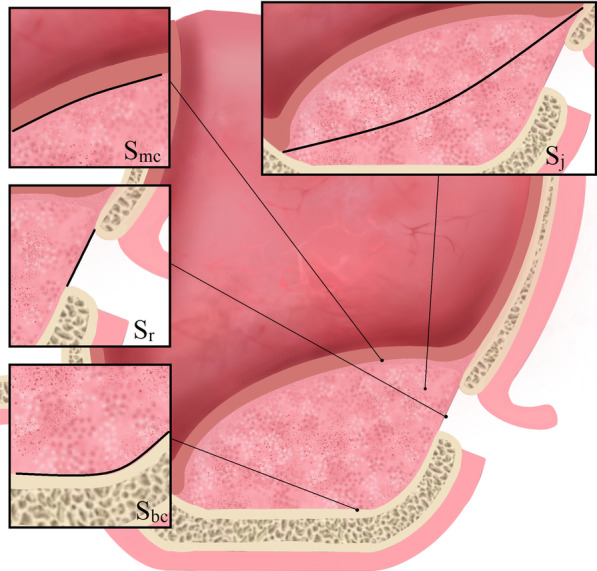
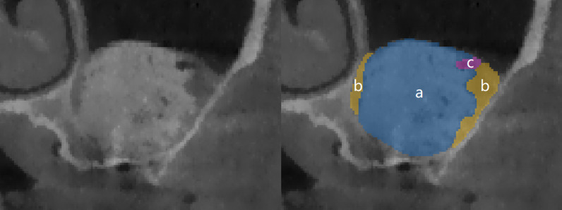
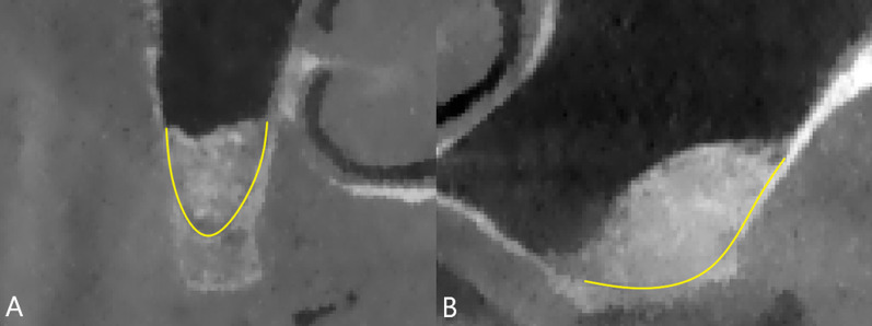
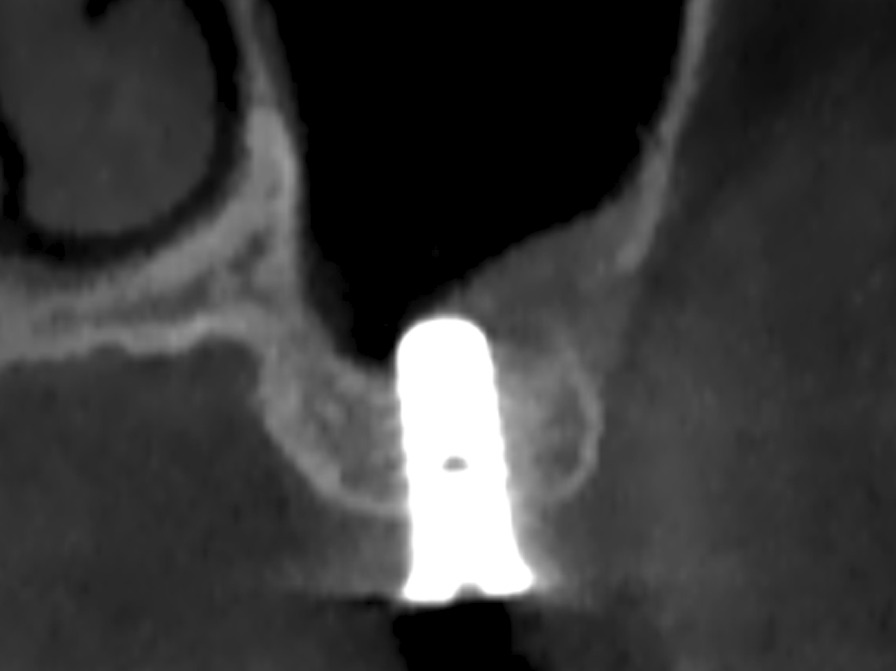
Methods: Cone beam computed tomography (CBCT) images were collected of patients requiring maxillary sinus floor elevation (residual bone height < 6 mm) for standard-length implant placement before surgery, after surgery, and at 6-month follow-up visits. The postoperative sub-antral space volume (V1) and surface area (S1), and the remaining volume after six months of healing (V2) were measured. Then, the contact area of sub-antral space with maxillary sinus bone (Sbc) and the Schneiderian membrane (Smc), the absorbed volume during healing (Va), and the percentage of remaining volume (V2%) and absorbed volume (Va%) were calculated. The correlation between anatomical parameters was analyzed using multiple linear regression.
Results: A total of 62 maxillary sinuses from 56 patients were augmented, of which 57 were considered for the final analysis (5 withdrew due to perforation). Multiple linear regression results demonstrated that Sbc was significantly positively correlated with Va (β coefficient = 0.141, p < 0.01) without correlation between Smc and Va (β coefficient = - 0.046, p = 0.470). There was a positive correlation between Sbc and V2% (β coefficient = 2.269, p < 0.05).
Conclusions: This study confirmed that the size of the Sbc in lateral window sinus elevation surgery affected osteogenesis after six months of healing. Clinicians should assess the sinus contour type preoperatively, then consider whether it is necessary to expand the range of the Schneiderian membrane elevation to avoid excessive collapse of the sub-antral space.
Trial registration: Chinese Clinical Trial Registry (ChiCTR), ChiCTR2200057924. Registered 22 March 2022-Retrospectively registered.
Keywords: Bone regeneration; CT imaging; Clinical research; Maxillary sinus; Sinus floor elevation.
© 2022. The Author(s).
Conflict of interest statement
All authors declare that they have no competing interests.
Figures
Similar articles
-
Nongrafted sinus floor elevation with a space-maintaining titanium mesh: case-series study on four patients.Clin Implant Dent Relat Res. 2014 Dec;16(6):893-903. doi: 10.1111/cid.12064. Epub 2013 Mar 28. Clin Implant Dent Relat Res. 2014. PMID: 23551586
-
Radiographic Outcomes of Transcrestal Sinus Floor Elevation With RBH ≤ 5 mm: Non-Perforation and Laterally Repaired Cases.Clin Implant Dent Relat Res. 2025 Apr;27(2):e70034. doi: 10.1111/cid.70034. Clin Implant Dent Relat Res. 2025. PMID: 40197860
-
Three-dimensional changes and influencing factors of tent space following osteotome sinus floor elevation without grafting: A 48-month retrospective radiographic study.Clin Oral Implants Res. 2024 Oct;35(10):1251-1261. doi: 10.1111/clr.14312. Epub 2024 Jun 14. Clin Oral Implants Res. 2024. PMID: 38873850
-
[Surgical dilemmas. Sinus floor elevation].Ned Tijdschr Tandheelkd. 2008 Dec;115(12):668-72. Ned Tijdschr Tandheelkd. 2008. PMID: 19149134 Review. Dutch.
-
Anatomy of the Maxillary Sinus and the Role of CT Scans in Maxillary Sinus Augmentation Surgery.Clin Implant Dent Relat Res. 2025 Apr;27(2):e70019. doi: 10.1111/cid.70019. Clin Implant Dent Relat Res. 2025. PMID: 40197815 Review.
References
Publication types
MeSH terms
Grants and funding
LinkOut - more resources
Full Text Sources
