. 2022 Dec 16:33:101413.
doi: 10.1016/j.bbrep.2022.101413. eCollection 2023 Mar.

Affinity purification of serum-derived anti-IA-2 autoantibodies in type 1 diabetes using a novel MBP-IA-2 fusion protein

Affiliations

Affinity purification of serum-derived anti-IA-2 autoantibodies in type 1 diabetes using a novel MBP-IA-2 fusion protein

Thilini Mendis et al. Biochem Biophys Rep. .

Abstract

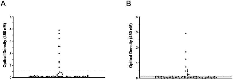
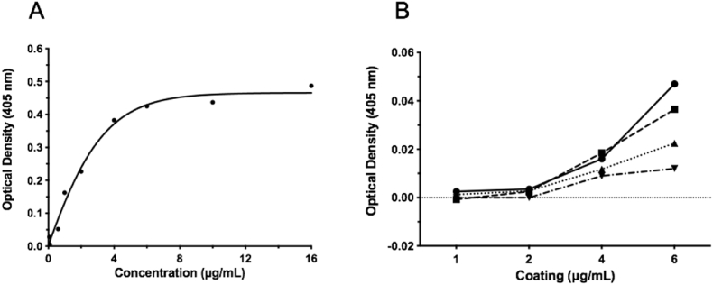
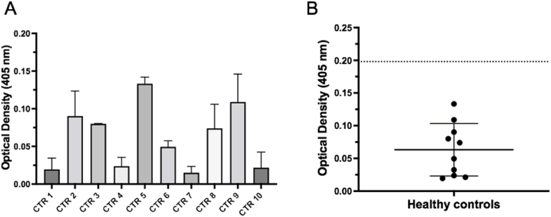
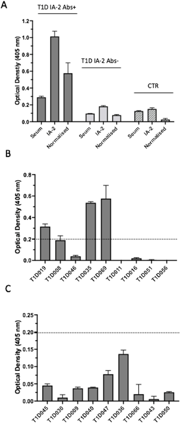
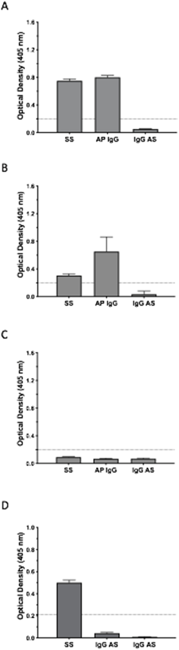
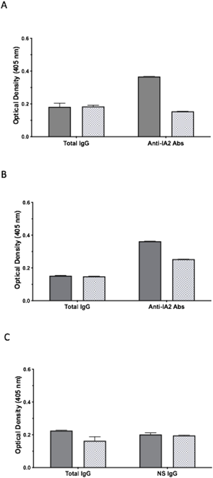
Autoantibodies targeting epitopes contained within the intracellular domain (IC) of the protein phosphatase-like islet antigen 2 (IA-2) are a common marker of autoimmune type 1 diabetes (T1D), however the isolation of genuine, serum derived anti-IA-2 autoantibodies has proven challenging due to a lack of suitable bioassays. In the current study, an ELISA format was developed for affinity purification of human anti-IA-2ic autoantibodies utilizing a fusion protein (FP) incorporating maltose binding protein and the full-length IA-2IC domain. Using a T1D patient cohort validated for anti-IA-2ic autoantibodies by commercial ELISA, we demonstrate the MBP-IA-2ic FP ELISA detects serum anti-IA-2IC autoantibodies from 3 of 9 IA-2 positive patients. Further to this, a multi-plate MBP-IA-2ic FP ELISA protocol specifically affinity purifies IgG enriched for anti-IA-2ic autoantibodies. Interestingly, serum derived autoantibodies immobilised on the MBP-IA-2ic FP ELISA demonstrate increased Kappa light chain usage when compared to the respective total IgG derived from donor patients, suggesting a clonally restricted repertoire of anti-IA-2ic autoantigen specific B plasma cells is responsible for autoantibodies detect by the MBP-IA-2ic FP ELISA. This study is the first to demonstrate the generation of specific, genuine human derived anti-IA-2ic autoantibodies, thereby facilitating further investigation into the origin and functional significance of IA-2 autoantibodies in T1D.

Keywords: Affinity purification; Autoantibodies; Clonal restriction; FP, fusion protein; GAD65, glutamic acid decarboxylase 65; IA-2; IA-2, islet antigen 2; IC, intracellular; OD, optical density; T1D, Type 1 Diabetes; Type 1 diabetes; β, beta cell.


Conflict of interest statement

The authors declare that they have no known competing financial interests or personal relationships that could have appeared to influence the work reported in this paper.

Figures

Fig. 1
Fig. 1
Prevalence of anti-IA-2 and ZnT8 autoantibodies in a cohort of 66 patients with established type 1 diabetes. Patients with confirmed type 1 diabetes were assessed for autoantibodies against the intracellular domain of IA-2 (A), and ZnT8 (B) using commercial ELISA assays (RSR Limited, Cardiff UK). Antibodies to IA-2 were present in individual serum samples from 9 of 66 patients, with individual serum samples from 10 patients containing anti-ZnT8 autoantibodies. Serum samples were assessed in triplicate. Assay cut offs for positivity were determined by standards and controls as per manufacturers instructions, and indicated on the figures by dotted lines. Individual points represent mean OD values for individual patient serum samples.
Fig. 2
Fig. 2
Optimisation of coating concentration and serum dilution in an MBP-IA-2 fusion protein ELISA. A. Incubation of ELISA plates with between 0.06 and 16 μg/mL with MPB-IA-2 fusion protein resulted in saturation at 6 μg/mL as determined using an anti-MBP monoclonal antibody. B. Subsequently, fusion protein coated ELISA plates were incubated with serum from 2 healthy individuals diluted at 1:10 (solid line - circle), 1:20 (dashed line - square), 1:50 (dotted line - triangle) and 1:100 (dotted/dashed line - inverted triangle). Background binding was highest at a coating concentration of 6 μg/mL, followed by 4 μg/mL. At most coating concentrations, serum dilutions of 1:10 produced the highest background and 1:100 the lowest. To achieve maximum assay sensitivity and acceptable background serum binding, a coating concentration of 4 μg/mL with serum dilution of 1:20 was selected for all subsequent fusion proten ELISAs. Data points represent the mean of individual experimental conditions. All experimental conditions were performed in triplicate.
Fig. 3
Fig. 3
Determination of background absorption of serum from healthy controls in a MBP-IA-2 fusion protein ELISA. A. Absorption values of serum (1:20 dilution) from 10 individual healthy donors (CTR) ranged from approximately 0.03 to 0.13 at a wavelength of 405 nM at a MBP-IA-2 fusion protein concentration of 4 μg/mL. B. Following the ELISA, the means from the 10 individual CTR serum samples were plotted and overall CTR binding means and SD assessed. As demonstrated by the Scatter Plot, the combined mean of the 10 CTR samples (solid long bar) was 0.063, with a SD ( ± solid short bars) of 0.045. Using the CTR data to set the value for positivity for anti-IA-2 autoantibodies, a value equal to the combined mean plus 3 SDs (dotted line) was generated, which provided a cut off absorbance of 0.198. This value was used in subsequent ELISAs as a cut off for the presence of anti-IA-2 autoantibodies in serum samples from patients with Type 1 diabetes. Data for panel A represents normalised means and SEM from at least n = 6 individual replicates from 2 separate experiments, with the means (solid circles) used to generate the scatter plot in panel B.
Fig. 4
Fig. 4
Detection of anti-IA-2 autoantibodies in the serum of a subset of IA-2 positive type 1 diabetes patients using an MBP-IA-2 fusion protein (FP) ELISA. A. Representative MBP-IA-2 fusion protein ELISA binding using patient and control serum. Serum from a patient positive (T1D IA-2+) and negative (T1D IA-2-) for IA-2 autoantibodies by RSR commercial ELISA were assessed by MBP-IA-2 FP ELISA. Normalised OD values for the anti-IA-2 antibody positive patient of 0.58 exceeded the 0.198 cut off for positivity by MBP-IA-2 FP ELISA, as determined in Fig. 3. By comparison, the normalised OD of 0.078 for the T1D IA-2 – patient was below the cut off, as was value (0.026) for the healthy control serum (CTR). B. Nine individual sera from T1D patients positive for anti-IA-2 autoantibodies by RSR ELISA were then screened using the MPB-IA-2 FP ELISA. Of these, 3 (T1D019, T1D035 and T1D069) resulted in OD values above the assay cut off for positivity (dotted line). C. By comparison to the RSR positive patients, all 9 sera from RSR negative patients produced OD values by MBP-IA-2 ELISA below the assay cut off for positivity (dotted line). Values represent mean +SEM of at least n = 6 individual replicates. Each serum was assessed by MBP-IA-2 ELISA on at least 2 separate occasions.
Fig. 5
Fig. 5
Confirmation of reactivity and specificity in anti-IA-2 autoantibodies affinity purified using the MBP-IA-2 fusion protein ELISA. A. Multi-plate ELISA capture and subsequent elution of anti-IA-2 immunoglobulin (AP IgG) from patient T1D069 achieved OD values above the cut off for anti-IA-2 positivity (dotted line) on the MBP-IA-2 FP ELISA. Similar positive OD binding was achieved with the starting serum (SS) from the same patient. By contrast, when the starting serum was diluted to match the IgG concentration of the AP IgG, positivity on the ELISA was lost (IgG AS). B. Similar to T1D069, both SS and AP IgG samples from patient T1D019 achieved positivity on the MBP-IA-2 FP ELISA, with positivity lost in the IgG AS sample. C. No positivity was achieved on the ELISA by SS, AP IgG or IgG AS samples from a healthy control. D. To assess specificity of the affinity purified anti-MBP-IA-2 FP autoantibodies, SS, IgG AS and IgG AS samples from patient T1D069 were subjected to MBP-ZnT8 fusion protein ELISA. As expected, the starting serum of this patient achieved positivity on the MBP-ZnT8 fusion protein ELISA (this patient tested positive for both anti-IA-2 and ZnT8 antibodies by RSR ELISA). However, consistent with specific purification of anti-IA-2 antibodies, positivity on the MBP-ZnT8 fusion protein ELISA was lost in the AP IgG sample generated by multi-plate MBP-IA-2 fusion protein ELISA capture. As expected, the IgG AS sample from this patient was also negative on the MBP-ZnT8 FP ELISA. Data represents normalised means and SEM from at least n = 6 individual replicates from 2 separate experiments.
Fig. 6
Fig. 6
Predominance of Kappa light chains in anti-IA-2 autoantibodies suggest clonal restriction of autoreactive B cells. A. Assessment of clonality in anti-IA-2 autoantibodies was achieved by determination of the Kappa/Lambda light chain ratio in immobilised total IgG (Total IgG) verses anti-MPB-IA-2 fusion protein specific IgG (Anti-IA-2 Abs) from a patient (T1D069) positive for anti-IA-2 autoantibodies. While the Kappa/Lambda ratio of approximately 1:1 in total IgG suggests near equal light chain representation in total serum IgG, for antibodies recognising IA-2 epitopes present in the MBP-IA-2 fusion protein, a ratio of approximately 2:1 indicates predominantly Kappa chains, indicative of clonal restriction in anti-IA-2 autoreactive B plasma cells. B. A predominance of Kappa chains in anti-IA-2 autoantibodies (Kappa/Lambda ratio 1.44:1) relative to total IgG (Kappa/Lambda ratio 1:1) in a second patient (T1D019) positive by MPB-IA-2 fusion protein ELISA confirms clonal restriction of anti-IA-2 autoreactive B plasma cells. C. In contrast to patients with T1D, the Kappa/Lambda ratio of IgG from a healthy control (CTR068) did not display evidence of increased Kappa expression in antibodies immobilised by fusion protein ELISA. The Kappa/Lambda ratio of 1.36:1 was observed in total IgG from this individual, with a post MBP-IA-2 ELISA (NS IgG) ratio of 1.03:1. Data represents normalised means and SEM from at least n = 6 individual replicates from 2 separate experiments.

References

    1. Atkinson M.A., Eisenbarth G.S., Michels A.W. Type 1 diabetes. Lancet. 2014;383:69–82. - PMC - PubMed
    1. Michels A., Zhang L., Khadra A., Kushner J.A., Redondo M.J., Pietropaolo M. Prediction and prevention of type 1 diabetes: update on success of prediction and struggles at prevention. Pediatr. Diabetes. 2015;16:465–484. - PMC - PubMed
    1. Regnell S.E., Lernmark A. Early prediction of autoimmune (type 1) diabetes. Diabetologia. 2017;60:1370–1381. - PMC - PubMed
    1. Mannering S.I., Pathiraja V., Kay T.W. The case for an autoimmune aetiology of type 1 diabetes. Clin. Exp. Immunol. 2016;183:8–15. - PMC - PubMed
    1. Willcox A., Richardson S.J., Bone A.J., Foulis A.K., Morgan N.G. Analysis of islet inflammation in human type 1 diabetes. Clin. Exp. Immunol. 2009;155:173–181. - PMC - PubMed

LinkOut - more resources