

High-dose steroid-responsive COVID-19-related encephalopathy with a sudden onset of dysarthria mimicking stroke: a case report
- PMID: 36579076
- PMCID: PMC9791002
- DOI: 10.1177/11795735221147218
High-dose steroid-responsive COVID-19-related encephalopathy with a sudden onset of dysarthria mimicking stroke: a case report
Abstract
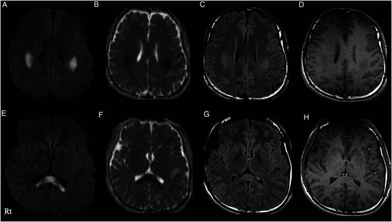
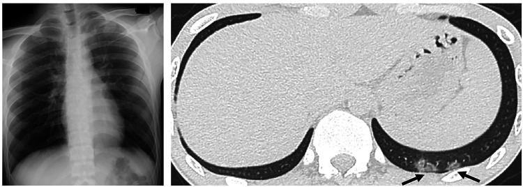
There has been limited research on encephalitis/encephalopathy, which is a less common coronavirus disease 2019 (COVID-19) neurological complication. The differentiation between stroke and encephalopathy with stroke mimickers is challenging in patients with COVID-19. Here, we describe a case of COVID-19-related encephalopathy mimicking stroke that was successfully treated with high-dose steroid pulse therapy. The patient suddenly experienced language disturbance with a left facial droop and symmetric numbness in his upper limbs. Magnetic resonance imaging (MRI) scans revealed hyperintensities in both the white matter and splenium. No pneumonia was observed. MRI abnormalities and neurological symptoms resolved after steroid pulse therapy and administration of remdesivir. High-dose steroid pulse treatment (for 3 days) might alleviate COVID-19-related encephalopathy.
Keywords: COVID-19; Encephalitis; coronavirus; steroid; stroke.
© The Author(s) 2022.
Conflict of interest statement
The author(s) declared no potential conflicts of interest with respect to the research, authorship, and/or publication of this article.
Figures
Similar articles
-
Mild Encephalopathy with Reversible Lesions in the Splenium of Corpus Callosum and Bilateral Cerebral Deep White Matter in Identical Twins.Pediatr Rep. 2016 Sep 19;8(3):6615. doi: 10.4081/pr.2016.6615. eCollection 2016 Sep 19. Pediatr Rep. 2016. PMID: 27777703 Free PMC article.
-
Case Report: Early-Onset Charcot-Marie-Tooth 2N With Reversible White Matter Lesions Repeatedly Mimicked Stroke or Encephalitis.Front Pediatr. 2022 Jul 13;10:935721. doi: 10.3389/fped.2022.935721. eCollection 2022. Front Pediatr. 2022. PMID: 35911843 Free PMC article.
-
Encephalopathy in COVID-19 Presenting With Acute Aphasia Mimicking Stroke.Front Neurol. 2020 Oct 19;11:587226. doi: 10.3389/fneur.2020.587226. eCollection 2020. Front Neurol. 2020. PMID: 33193051 Free PMC article.
-
Hashimoto Encephalopathy Presenting With Stroke-Like Episodes in an Adolescent Female: A Case Report and Literature Review.Pediatr Neurol. 2016 Jun;59:62-70. doi: 10.1016/j.pediatrneurol.2016.02.003. Epub 2016 Mar 3. Pediatr Neurol. 2016. PMID: 27033176 Review.
-
Neurological Sequelae of COVID-19.J Integr Neurosci. 2022 Apr 6;21(3):77. doi: 10.31083/j.jin2103077. J Integr Neurosci. 2022. PMID: 35633158 Review.
Cited by
-
The youngest infant with COVID-19-associated necrotizing encephalitis in Asia: A case report.SAGE Open Med Case Rep. 2023 Nov 20;11:2050313X231211713. doi: 10.1177/2050313X231211713. eCollection 2023. SAGE Open Med Case Rep. 2023. PMID: 38022854 Free PMC article.
-
Unlocking the code for stroke treatment and care.J Cent Nerv Syst Dis. 2024 Sep 3;16:11795735241280805. doi: 10.1177/11795735241280805. eCollection 2024. J Cent Nerv Syst Dis. 2024. PMID: 39238575 Free PMC article. No abstract available.
References
Publication types
LinkOut - more resources
Full Text Sources
