. 2023 Feb;10(2):276-291.
doi: 10.1002/acn3.51719. Epub 2022 Dec 28.

Hyperacute immune responses associate with immediate neuropathology and motor dysfunction in large vessel occlusions

Affiliations

Hyperacute immune responses associate with immediate neuropathology and motor dysfunction in large vessel occlusions

Mudassir Farooqui et al. Ann Clin Transl Neurol. 2023 Feb.

Abstract

Objective: Despite successful endovascular therapy, a proportion of stroke patients exhibit long-term functional decline, regardless of the cortical reperfusion. Our objective was to evaluate the early activation of the adaptive immune response and its impact on neurological recovery in patients with large vessel occlusion (LVO).

Methods: Nineteen (13 females, 6 males) patients with acute LVO were enrolled in a single-arm prospective cohort study. During endovascular therapy (EVT), blood samples were collected from pre and post-occlusion, distal femoral artery, and median cubital vein (controls). Cytokines, chemokines, cellular and functional profiles were evaluated with immediate and follow-up clinical and radiographic parameters, including cognitive performance and functional recovery.

Results: In the hyperacute phase (within hours), adaptive immune activation was observed in the post-occlusion intra-arterial environment (post). Ischemic vascular tissue had a significant increase in T-cell-related cytokines, including IFN-γ and MMP-9, while GM-CSF, IL-17, TNF-α, IL-6, MIP-1a, and MIP-1b were decreased. Cellularity analysis revealed an increase in inflammatory IL-17+ and GM-CSF+ helper T-cells, while natural killer (NK), monocytes and B-cells were decreased. A correlation was observed between hypoperfused tissue, infarct volume, inflammatory helper, and cytotoxic T-cells. Moreover, helper and cytotoxic T-cells were also significantly increased in patients with improved motor function at 3 months.

Interpretation: We provide evidence of the activation of the inflammatory adaptive immune response during the hyperacute phase and the association of pro-inflammatory cytokines with greater ischemic tissue and worsening recovery after successful reperfusion. Further characterization of these immune pathways is warranted to test selective immunomodulators during the early stages of stroke rehabilitation.


Conflict of interest statement

The authors have no financial conflicts of interest relevant to this study.

Figures
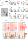
Figure 1
Figure 1
Altered immune microenvironment within hours after stroke onset. We quantified 22 cytokines, growth factors, and cerebrovascular injury‐related proteins in the plasma of hyper‐acute stroke patients. Arterial plasma was isolated after an average of 7.5 h post‐ischemic injury downstream of clot (Post), upstream of clot (Pre), and distant from clot (IA). Representative angiography of an M1‐MCA occlusion (red arrow). The location of pre‐clot arterial sampling (yellow arrowhead) and post‐clot arterial sampling (orange arrowhead) is shown (A). Post‐clot arterial sampling is not visible due to angiography methodology. In (B–F), plasma proteins were quantified using the Bioplex Immunotherapy Assay or ELISA‐based quantification. Inflammation‐related protein, IFN‐γ was significantly increased in post‐clot plasma, relative to the intra‐arterial control site. Inflammatory cytokines, GM‐CSF, TNF‐α, IL‐17, and IL‐6 were decreased relative to the control site (B). Anti‐inflammatory proteins IL‐4 and IL‐5 are decreased in post‐ictus plasma (C). Pro‐survival IL‐2, IL‐7, and IL‐15 were significantly decreased relative to the control site (D). Macrophage‐associated proteins IP‐10, MIP‐1a, and MIP‐1b were also found to be significantly decreased relative to the control site. Vascular injury‐related protein MMP9 was found to be increased, while VEGF was decreased relative to the control site (E). Relative comparison of inflammatory and anti‐inflammatory cytokines in their respective locations (F). All samples were tested in duplicate and concomitantly assayed with manufacturer‐provided standards. Parametric data were graphed as violin plots with individual data points and analyzed by repeated measures one‐way ANOVA with Tukey's multiple comparisons test. A p‐value of ≤0.05 was considered significant. *p < 0.05; **p < 0.01; ***p < 0.001.
Figure 2
Figure 2
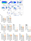
Decreased Monocytes, NK, and B‐cells distal to the clot. Immediately after sampling, blood was processed and stained, and major adaptive and innate immune cell subsets were quantified using flow cytometry. We qualified our flow data by using a gating strategy that focused on events within the laminar flow, removed doublets, and focused on live (GhostDye 780) cells (A). Blood isolated from the blood downstream of the clot (Post) was found to have a decrease in hematopoietic (CD45+) cells relative to blood upstream of the clot (B). Within the innate immune cell populations, only natural killer (CD161+), and monocytes (CD11b+) cells were found to be decreased in number (C). Within the adaptive immune cell populations, only B‐cells (CD19+) were found to be decreased in number (D). All three sample sites of each patient were assayed together immediately after the acquisition of blood. Nonparametric data graphed as median with range and analyzed using Friedman's test with Dunn's multiple comparison test correction. A p‐value of ≤0.05 was considered significant. *p < 0.05; **p < 0.01; ***p < 0.001.
Figure 3
Figure 3
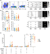
Increase in multiple pro‐inflammatory helper T‐cells in hyperacute post‐occlusion blood. Immediately following blood processing, intracellular cytokine production was measured by labeling with fluorescent antibodies specific to indicated protein and quantified using flow cytometry. Representative dot plots indicating the intensity of cytokine levels for GM‐CSF, IFN‐γ, IL‐17, and IL‐10 are shown (A). Single‐dimensional analysis revealed an increase in GM‐CSF+ CD4 and IL‐17+ CD4 T‐cells in the post‐occlusion samples (B). Two‐dimensional analysis reveals an increase in IFN‐γ+GM‐CSF+ CD4 T‐cells in the post‐occlusion samples (C). Three‐dimensional analysis reveals an increase in GM‐CSF+IFN‐γ+IL‐17+ and GM‐CSF+IFN‐γ+IL‐10+ CD4 T‐cells in the post‐occlusion samples (D). All three sample sites of each patient were assayed together immediately after the acquisition of blood. Parametric data were graphed as mean ± SD and analyzed by repeated measures one‐way ANOVA, Tukey's multiple comparisons test.
Figure 4
Figure 4
Clinically relevant immune correlates in hyper‐acute stroke patients. Correlation analysis between biological (cellularity, cytokine, and function profiles) and clinical (perfusion, infarct volume, cognitive and motor function) parameters were performed. (A) Representative neuroimaging of patients with small (left column) and large (right column) ischemic strokes. Perfusion imaging showing CBF < 30% (top row) and T max >6 s (second row); and DVI‐MRI (bottom row) after EVT is shown. Tissue damage‐associated proteins (MCP‐1) located in the femoral arterial sample (IA) were found to correlate with perfusion negatively (B top row), while RANTES were associated with motor function at 3 months (B, bottom row). B‐cell cellularity was found to correlate with perfusion in the intra‐arterial and post‐occlusion locations positively (C). Post‐occlusion inflammatory CD4 T‐cells were found to correlate with T max 6 s perfusion negatively and femoral arterial samples correlate with infarct volume positively (D). Pre‐occlusion inflammatory CD8 T‐cells were found to correlate with infarct volume (E). Statistical analysis for (B–E) was performed using the LASSO method, and an r‐value of >0.7 and p‐value ≤0.05 was considered significant.
Figure 5
Figure 5
Decreased adaptive cellularity and increased plasma neurotoxicity during hyperacute stroke associates with poor long‐term recovery. At 3 months, motor function recovery is distinct and stratified into those whose performance declines as revealed by an increase in mRS scores (Declined), those who maintain function and no change in mRS scores (No change), and those who improve and had a decrease in mRS scores (Improved, A). Motor function‐dependent separation of patients reveals an increase in pre‐occlusion IL‐15 and a decrease in post‐occlusion IP‐10 in patients with poor functional recovery (B). Conversely, improving patients have an increased quantity of GM‐CSF+IL‐17+ CD4 T‐cells in the post‐occlusion sample relative to the distal control sample (IA), which is absent in declining patients (C). Declining patients exhibit a loss of both helper and cytotoxic T‐cell cellularity in the distal arterial sample, as compared to recovering patients (D). After in vitro culture with patient plasma (5%) from declining patients, murine brain cultures exhibit a decrease in viable neurons (NeuN+DAPI+, arrows) as compared to brain cells cultured with the plasma of improving patients. Representative immunofluorescence image (left) and cumulative data (right). Quantification was assessed per well using four random independent images averaged for each patient sample site (E). Red = NeuN and blue = DAPI. Parametric data were graphed as mean ± SD and analyzed by repeated measures one‐way ANOVA, Tukey's multiple comparisons test.

References

    1. Zaidat OO, Fifi JT, Nanda A, et al. Endovascular treatment of acute ischemic stroke with the penumbra system in routine practice: COMPLETE registry results. Stroke. 2022;53(3):769‐778. - PMC - PubMed
    1. De Meyer SF, Stoll G, Wagner DD, Kleinschnitz C. von Willebrand factor: an emerging target in stroke therapy. Stroke. 2012;43(2):599‐606. - PMC - PubMed
    1. De Meyer SF, Denorme F, Langhauser F, Geuss E, Fluri F, Kleinschnitz C. Thromboinflammation in stroke brain damage. Stroke. 2016;47(4):1165‐1172. - PubMed
    1. Xing C, Arai K, Lo EH, Hommel M. Pathophysiologic cascades in ischemic stroke. Int J Stroke. 2012;7(5):378‐385. - PMC - PubMed
    1. Qin X, Akter F, Qin L, et al. Adaptive immunity regulation and cerebral ischemia. Front Immunol. 2020;11:689. - PMC - PubMed

Publication types

Substances

LinkOut - more resources