. 2023 Jan;27(2):246-258.
doi: 10.1111/jcmm.17645. Epub 2022 Dec 29.

Protective effects of activated vitamin D receptor on radiation-induced intestinal injury

Affiliations

Protective effects of activated vitamin D receptor on radiation-induced intestinal injury

Yuhan Lin et al. J Cell Mol Med. 2023 Jan.

Abstract

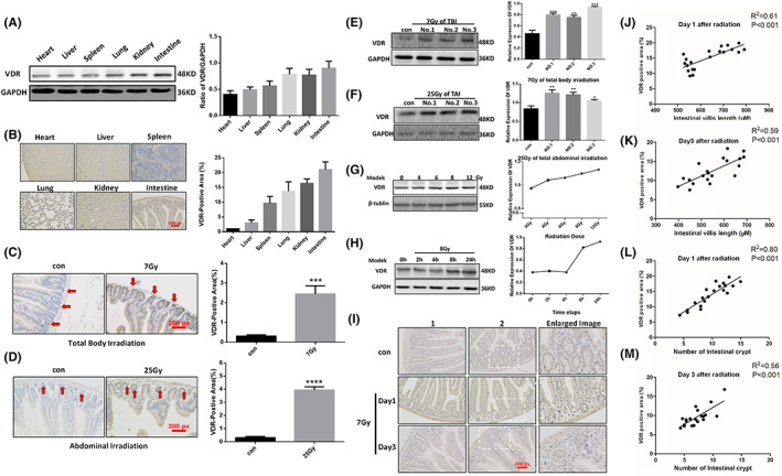
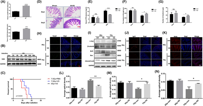
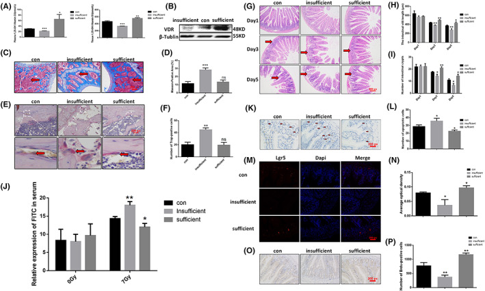
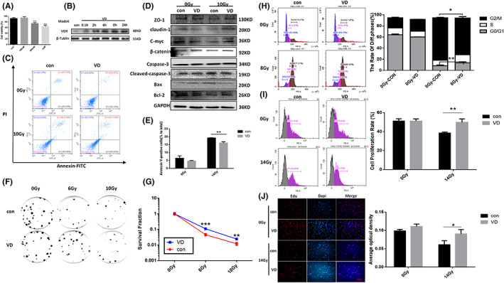
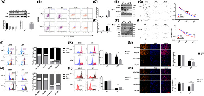
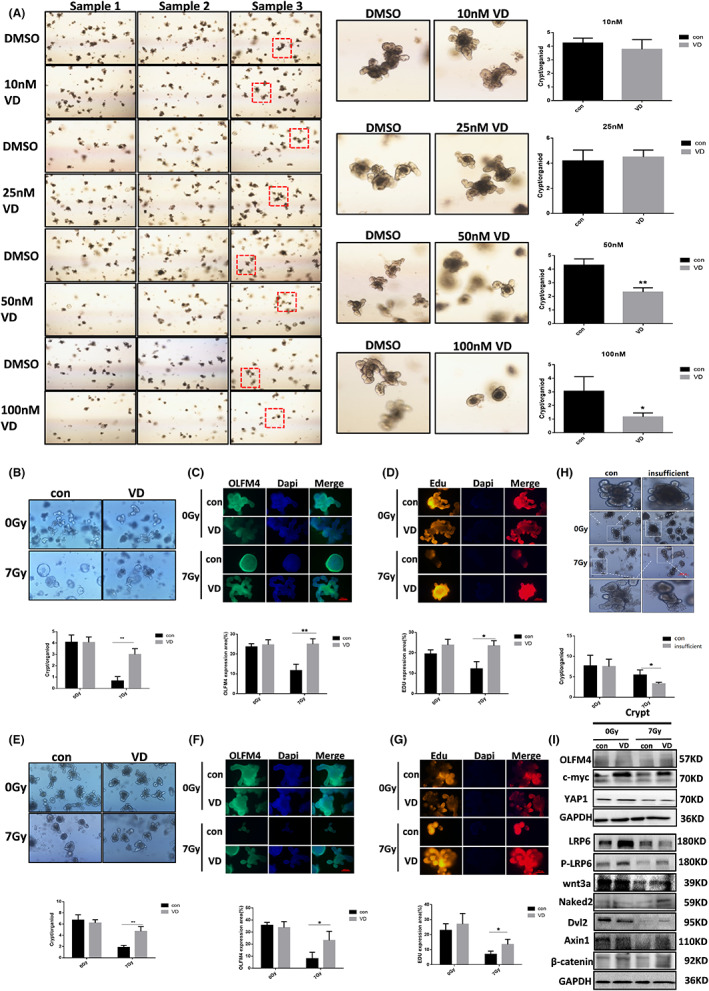
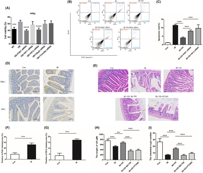
Radiation-induced intestinal injury (RIII) is a common complication after radiation therapy in patients with pelvic, abdominal, or retroperitoneal tumours. Recently, in the model of DSS (Dextran Sulfate Sodium Salt) -induced intestinal inflammatory injury, it has been found in the study that transgenic mice expressing hVDR in IEC (Intestinal Epithelial Cell) manifest highly anti-injury properties in colitis, suggesting that activated VDR in the epithelial cells of intestine may inhibit colitis by protecting the mucosal epithelial barrier. In this study, we investigated the effect of the expression and regulation of VDR on the protection of RIII, and the radiosensitivity in vitro experiments, and explored the initial mechanism of VDR in regulating radiosensitivity of IEC. As a result, we found that the expression of VDR in intestinal tissues and cells in mice can be induced by ionizing radiation. VDR agonists are able to prolong the average survival time of mice after radiation and reduce the radiation-induced intestinal injury. For lack of vitamin D, the radiosensitivity of intestinal epithelial cells in mice increased, which can be reduced by VDR activation. Ensuing VDR activation, the radiation-induced intestinal stem cells damage is decreased, and the regeneration and differentiation of intestinal stem cells is promoted as well. Finally, on the basis of sequencing analysis, we validated and found that VDR may target the HIF/PDK1 pathway to mitigate RIII. We concluded that agonism or upregulation of VDR expression attenuates radiation-induced intestinal damage in mice and promotes the repair of epithelial damage in intestinal stem cells.

Keywords: HIF/PDK1; radiation protection; radiation-induced intestinal injury; tumour radiotherapy; vitamin D.


Conflict of interest statement

The authors have no potential conflict of interests.

Figures

FIGURE 1
FIGURE 1
Effects of ionizing radiation on VDR expression in intestinal tissues and epithelial cells of mice. (A) VDR expression in various organs and tissues of mice was testified by Western Blot Analysis. (B) VDR expression in various organs of mice was detected by immunohistochemistry. (C) Immunohistochemical staining of VDR in intestinal tracts of mice after 7 Gy of general irradiation. (D) Immunohistochemical staining of VDR in intestinal tracts of mice after 25 Gy of abdominal irradiation. (E) VDR in intestinal tracts of mice was detected by Western Blot Analysis after 7 Gy of general irradiation. (F) VDR in intestinal tracts of mice was detected by Western Blot Analysis after 25 Gy of abdominal irradiation. Con stands for non‐irradiated group and the others are irradiated groups. No. 1–3 represented three separate samples. G. Radiation‐induced VDR expression in Modek cells was dose‐dependent. (H) VDR expression induced by 8 Gy irradiation in Modek cells was time‐dependent. (I) VDR immunohistochemical map. (J) The relationship between intestinal villus length and VDR expression on day 1 after irradiation. (K) The relationship between intestinal villus length and VDR expression on day 3 after irradiation. (L) The relationship between intestinal crypt number and VDR expression on day 1 after irradiation. (M) The relationship between intestinal crypt number and VDR expression on day 3 after irradiation. The statistical results are expressed by Mean ± SD, *** and **** represents p < 0.001 and p < 0.0001, respectively. Each experiment was repeated for three times. N = 7
FIGURE 2
FIGURE 2
VDR agonists alleviate radiation‐induced intestinal damage in mice. (A) Effect of VDR agonist on 1,25‐(OH)2D metabolism in mice. (B) Effect of VDR agonist on VDR expression in small intestines of mice. (C) VDR agonist prolonged the average survival time of mice after irradiation. (D) H&E pathological test on mouse intestine post irradiation. (E) Expression level of FITC‐dextran in mouse serum. (F) The length of villi. (G) The number of intestinal crypts in mice. (H) The effect of VDR agonists on radiation‐induced apoptosis of intestinal epithelial cells was detected by Tunel immunofluorescence assay. (I) The role Calcitriol (VD) played on apoptosis of intestinal tissue after irradiation. (J) The effect of Calcitriol (VD) on Lgr5 + ISC post irradiation was detected by Lgr5+ mRNA fluorescence in situ hybridization (FISH). (K) The effect of Calcitriol (VD) on the proliferative capability of intestinal cells after irradiation was detected by the BrdU assay. (L) The bar chart depicting TUNEL staining. (M) The bar chart depicting Lgr5+ mRNA fluorescence in situ hybridization. (N) The bar chart depicting the BrdU staining. The statistical results are expressed by Mean ± SD, *, *** and **** represents p < 0.05, p < 0.001 and p < 0.0001, respectively. Each experiment was repeated for three times. N = 10
FIGURE 3
FIGURE 3
Vitamin D insufficient/sufficient mouse model was constructed and radiation‐induced intestinal injury was analysed. (A) The level of 1,25‐(OH)2D in serum and intestinal tissues of Vitamin D insufficient/sufficient mice was detected by ELISA. (B) Intestinal VDR expression of Vitamin D insufficient/sufficient mice were detected by the Western blot test. (C) Femurs of Vitamin D insufficient/sufficient mice were stained by Masson's trichrome. (D) The bar chart depicting Masson Staining. (E) TRAP staining of femurs of Vitamin D insufficient/sufficient mice. (F). statistical chart depicting TRAP staining. (J) FITC‐dextran level in serum of Vitamin D insufficient/sufficient mice before and after irradiation. (G) H&E pathological test on the intestine of Vitamin D insufficient/sufficient mice post irradiation. (H) The length of villi stained with H&E. (I) The intestinal crypt number stained with H&E. (K) Immunohistochemical staining of Vitamin D insufficient/sufficient mice post irradiation. (L) Bar chart depicting the Immunohistochemical staining. (M) Mouse intestinal tissue was detected by Lgr5+ mRNA fluorescence in situ hybridization on day 3 after irradiation. (N) The statistical chart of fluorescence in situ hybridization. (O) Intestinal tissue of Vitamin D insufficient/sufficient mice was detected on day 3 after irradiation by the BrdU assay. (P) The bar chart depicting the BrdU staining. The statistical results are expressed by Mean ± SD, *, **, and *** represents p < 0.05, p < 0.01 and p < 0.001, respectively. Each experiment was repeated for three times. N = 10
FIGURE 4
FIGURE 4
VDR agonists lowered the radiosensitivity of Modek cells. A. The optimal concentration of VDR agonists was screened by CCK8 assay. (B) Calcitriol (VD) induced VDR expression in intestinal epithelial cells. (C) The effect of Calcitriol (VD) on the intestinal epithelial cell apoptosis post irradiation was detected by flow cytometry. (D) Western blot was used to verify the effect of Calcitriol (VD) on the expression of apoptosis protein, barrier protein and prolife in in intestinal epithelial cells after irradiation was verified by Western Blot Analysis. (E) The bar chart depicting apoptosis. (F) The effect of Calcitriol (VD) on the proliferative capability of Modek cells post irradiation was detected by clone formation assay. (G) The statistical chart of clone formation assay. (H) The effect of Calcitriol (VD) on the cell cycle of Modek cells post irradiation was detected by flow cytometry. (I) The effect of Calcitriol (VD) on the proliferative capability of Modek cells post irradiation was detected through Edu assay by flow cytometry. (J) The effect of Calcitriol (VD) on the proliferative capability of Modek cells post irradiation was detected through Edu assay by immunofluorescence. The statistical results are expressed by Mean ± SD, *, **, and *** represents p < 0.05, p < 0.01 and p < 0.001, respectively. Each experiment was repeated for three times.
FIGURE 5
FIGURE 5
The effect of overexpression/knockdown of VDR on the radiosensitivity of Modek cells. (A) The expression level of VDR in Modek cell lines was verified by Western blot/RT‐QPCR test. (B) The effect of knockdown/overexpression of VDR on intestinal epithelial cell apoptosis after irradiation was detected by flow cytometry assay. (C) The statistical chart depicting the effect of VDR knockdown on cell apoptosis after irradiation. (D) The statistical chart depicting the effect of VDR overexpression on cell apoptosis after irradiation. (E) The effect of VDR knockdown on the expression of corresponding proteins in intestinal epithelial cells after irradiation was verified by Western blot Analysis. (F) The effect of VDR overexpression on the expression of corresponding proteins in intestinal epithelial cells after irradiation was verified by Western blot Analysis. (G) The effect of VDR knockdown on the proliferative capability of Modek cells post irradiation was detected by clone formation assay. (H) The effect of VDR overexpression on the proliferative capability of Modek cells post irradiation was detected by clone formation assay. (I) The effect of VDR knockdown on the cell cycle of Modek cells post irradiation was detected by flow cytometry. (J) The effect of VDR overexpression on the cell cycle of Modek cells post irradiation was detected by flow cytometry. (K) The effect of VDR knockdown on the proliferative capability of Modek cells post irradiation was detected through Edu assay by flow cytometry. (L) The effect of VDR overexpression on the proliferative capability of Modek cells post irradiation was detected through Edu assay by flow cytometry. (M) The effect of VDR knockdown on the proliferative capability of Modek cells post irradiation was detected through Edu assay by immunofluorescence. (N) The effect of VDR overexpression on the proliferative capability of Modek cells post irradiation was detected through Edu assay by immunofluorescence. The statistical results are expressed by Mean ± SD, *, **, and *** represents p < 0.05, p < 0.01 and p < 0.001, respectively. Each experiment was repeated for three times.
FIGURE 6
FIGURE 6
Effect of VDR on the function of intestinal stem cells. (A) Drug toxicity testing on intestinal organoids. In vivo experiments: (B) Intestinal organoid culture. (C) Immunofluorescence staining of OLFM4. (D) Organoid proliferation was detected through Edu assay by immunofluorescence. In vitro experiments: (E) Intestinal organoid culture. (F) Immunofluorescence staining of OLFM4. (G) Organoid proliferation was detected through Edu assay by immunofluorescence. (H) Vitamin D deficiency aggravates damage to radiation‐induced intestinal organoids. (I) VDR agonists regulates Wnt pathway in intestinal epithelial cells after irradiation. The statistical results are expressed by Mean ± SD, *, and ** represents p < 0.05 and p < 0.01, respectively. Each experiment was repeated for three times.
FIGURE 7
FIGURE 7
VDR reduced the radiosensitivity of intestinal epithelial cells and alleviated radiation‐induced intestinal damage by targeting HIF/PDK1. (A) CCK8 assay verified the effect of each siRNA on the viability of Modek‐OE cells after irradiation. (B) With Pdk1 and Hif‐2 siRNA, the apoptosis was detected after irradiation and VDR agonist, 1,25‐(OH)2D using. (C) The quantity analysis for apoptosis in radiated Modek cells. (D) 3 days after irradiation, the expression of PDK1 and HIF‐2 were tested by immunohistochemical staining and were quantified (F, G). (E) Mice were intraperitoneal injected with Bx‐795 (inhibitor of PDK1) at 5 mg/kg and PT2385 (inhibitor of HIF2) at 10 mg/kg before irradiation and 1,25‐(OH)2D using, and 3 days later, the small intestine was taken to perform H&E staining, and the length of villi (H) and number of intestinal crypt (I) were quantified to indicate the degree of RIII. The statistical results are expressed by Mean ± SD, **, and *** represents p < 0.01 and p < 0.001, respectively. Each experiment was repeated for three times.

Similar articles

Cited by

References

    1. Lu L, Li W, Chen L, et al. Radiation‐induced intestinal damage: latest molecular and clinical developments. Future Oncol. 2019;15(35):4105‐4118. - PubMed
    1. Nussbaum ML, Campana TJ, Weese JL. Radiation‐induced intestinal injury. Clin Plast Surg. 1993;20(3):573‐580. - PubMed
    1. Cheng Y, du J, Liu R, et al. Novel chimeric TLR2/NOD2 agonist CL429 exhibited significant radioprotective effects in mice. J Cell Mol Med. 2021;25(8):3785‐3792. - PMC - PubMed
    1. An N, Liu T, Zhu B, et al. A bidirectional effect of Rac1 inhibition‐protects radiation‐induced intestinal injury while inhibits tumor. Life Sci. 2020;240:117105. - PubMed
    1. Xie LW, Cai S, Zhao TS, Li M, Tian Y. Green tea derivative (−)‐epigallocatechin‐3‐gallate (EGCG) confers protection against ionizing radiation‐induced intestinal epithelial cell death both in vitro and in vivo. Free Radic Biol Med. 2020;161:175‐186. - PubMed

Publication types