. 2023 Jan 1;16(1):dmm049699.
doi: 10.1242/dmm.049699. Epub 2023 Jan 16.

Induction of pancreatic neoplasia in the KRAS/TP53 Oncopig

Affiliations

Induction of pancreatic neoplasia in the KRAS/TP53 Oncopig

Pinaki Mondal et al. Dis Model Mech. .

Abstract

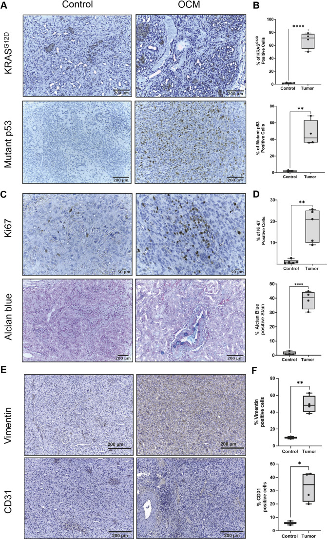
The 5-year survival of pancreatic cancer (PC) remains low. Murine models may not adequately mimic human PC and can be too small for medical device development. A large-animal PC model could address these issues. We induced and characterized pancreatic tumors in Oncopigs (transgenic swine containing KRASG12D and TP53R167H). The oncopigs underwent injection of adenovirus expressing Cre recombinase (AdCre) into one of the main pancreatic ducts. Resultant tumors were characterized by histology, cytokine expression, exome sequencing and transcriptome analysis. Ten of 14 Oncopigs (71%) had gross tumor within 3 weeks. At necropsy, all of these subjects had gastric outlet obstruction secondary to pancreatic tumor and phlegmon. Oncopigs with injections without Cre recombinase and wild-type pigs with AdCre injection did not show notable effect. Exome and transcriptome analysis of the porcine pancreatic tumors revealed similarity to the molecular signatures and pathways of human PC. Although further optimization and validation of this porcine PC model would be beneficial, it is anticipated that this model will be useful for focused research and development of diagnostic and therapeutic technologies for PC. This article has an associated First Person interview with the joint first authors of the paper.

Keywords: Oncopig; Pancreas; Pancreatic cancer; Porcine pancreatic cancer.


Conflict of interest statement

Competing interests The authors declare no competing or financial interests.

Figures

Fig. 1.
Fig. 1.
Porcine pancreatic anatomy and injection techniques. (A) Anterior aspect; top, cephalad. Double arrow and asterisk indicate the main pancreatic duct entering into duodenum via the pancreatic papilla (not visible). Arrows indicate the structure associated with each label. CA, celiac artery; CBD, common bile duct; CHA & PV, common hepatic artery and portal vein (not visible); CL, connecting lobe; D, duodenum; DL, duodenal lobe; E, esophagus; GA, gastric antrum; P, pylorus; PMV, portomesenteric vein emerging from underneath pancreas; SL, splenic lobe; SP, splenic pedicle. Inset: zoom out view. (B) Same specimen as panel A. Dotted yellow line boundaries indicate the pancreatic lobes. Yellow arrows indicate the approximate course of the pancreatic ductal system. Intersections of the DL, SL and CL form a continuous ductal loop; if interrupted, the system decompresses retrograde into the main pancreatic duct (PD; green arrow). (C) Same specimen, zoomed in. Yellow arrow indicates CL transection for ductal injection. Inset: CL has been transected; 22 g plastic catheter inserted (small white arrow) into CL duct (technique 2 for tumor induction). (D) Similar dissection in another subject, demonstrating circular DL–SL–CL lobar configuration. Inset: cannulization of transected CL duct with a 22 g plastic catheter. (E) Pancreatic parenchymal and main duct injection (technique 2). DL is inked (yellow arrows) for parenchymal adenovirus expressing Cre recombinase (AdCre) injection. Inset: PD accessed through a duodenotomy just opposite to the insertion of the PD; pancreatic papilla (1) then cannulated with 22 g catheter (2) for AdCre injection; DeBakey forceps (3) retracts duodenal wall. Large white arrows in A-E indicate cephalad.
Fig. 2.
Fig. 2.
Induction of Oncopig model (OCM) pancreatic tumor. (A) OCM necropsy <3 weeks after the tumor induction procedure; dashed yellow line indicates pancreatic phlegmon (contained tumor) at the location of AdCre injection. D, duodenum; Om, omentum. Scale, cm. (B) Another OCM necropsy <3 weeks after tumor induction procedure. Silk suture (double yellow arrow) is at the ligated proximal end of the connecting lobe (CL) at the intersection with the duodenal lobe (DL). Single yellow arrows indicate distal CL remnant (site of tumor). (C) Third OCM necropsy <3 weeks after tumor induction. Transection of CL phlegmon (yellow arrows) demonstrated firm nodular mass (tumor on pathology). Subject had typical gastric outlet obstruction (distended stomach). S, stomach; Sp, spleen. (D) Low-power view (H&E) of CL injection site (pancreatic phlegmon). Cords of inflammatory cells (yellow arrows) intermingled with hemorrhage (red arrows), with residual acini (black arrows). Scale bar: 500 µm. (E) Higher-power view of phlegmon from panel D. Sheets of tumor cells were apparent (white arrows), intermingled with residual acinar structures. Scale bar: 100 µm. (F) High-power view from panel E; individual tumor cells are indicated by white arrows. Double arrow indicates mitotic figure. Scale bar: 20 µm. Large white (A) and black (B,C) arrows indicate cephalad.
Fig. 3.
Fig. 3.
Immunohistochemistry of tumor-associated proteins and polysaccharide staining in pancreatic tumor from AdCre-induced Oncopigs versus pancreas from wild-type (WT) littermates. (A-F) Expression and quantification of KRAS (n=3, control and OCM) and p53 mutants (n=3, control and OCM) (A,B), Ki-67 (n=3, control and OCM) and Alcian Blue (n=3, control and OCM) (C,D), and vimentin (n=2, control and n=4, OCM) and CD31 (n=2, control and n=4, OCM) (E,F). Controls [WT pig with AdCre injection into the connecting lobe (same conditions as OCM treatment)] used for KRAS, mutant p53, Ki-67 and Alcian Blue were subjects 1083, 1087 and 1101. Controls for vimentin and CD31 were subjects 1087 and 1101. Unpaired two-tailed Student's t-test, *P<0.05, **P<0.01, ****P<0.0001.
Fig. 4.
Fig. 4.
Exome insertions and deletions in OCM tumors. All displayed maps compare exome sequence data between a group of four OCM pancreatic tumors (subjects 1090, 1092, 1095 and 1096 from Table 1) and normal porcine pancreas (i.e. Oncopig pancreas without AdCre injection); indels (insertions and deletions) refer to tumor exome with respect to normal. (A) Exome-wide map of all indels. (B) Indels predicted to have an effect on protein expression. (C) Indels involving key genes in human pancreatic cancer (PC). (D) Indels involving other genes related to human PC.
Fig. 5.
Fig. 5.
Transcriptomics of OCM pancreatic tumors versus WT pancreas. (A) Differential gene expression scatterplot (RNA sequencing) from WT pancreas (n=2) and pancreatic tumor (n=2). (B) Hierarchical cluster analysis of top 50 upregulated and downregulated genes. (C) KRAS and TP53 expression. (D) Enrichment (ES) score for Hallmark epithelial-to-mesenchymal transition (EMT) genes. FDR, false discovery rate; NES, normalized enrichment score. (E) Differential expression of selected EMT genes.

References

    1. Aiello, N. M., Maddipati, R., Norgard, R. J., Balli, D., Li, J., Yuan, S., Yamazoe, T., Black, T., Sahmoud, A., Furth, E. E.et al. (2018). EMT subtype influences epithelial plasticity and mode of cell migration. Dev. Cell 45, 681-695.e4. 10.1016/j.devcel.2018.05.027 - DOI - PMC - PubMed
    1. Begley, C. G. and Ellis, L. M. (2012). Drug development: raise standards for preclinical cancer research. Nature 483, 531-533. 10.1038/483531a - DOI - PubMed
    1. Bergeron, S., Lemieux, E., Durand, V., Cagnol, S., Carrier, J. C., Lussier, J. G., Boucher, M.-J. and Rivard, N. (2010). The serine protease inhibitor serpinE2 is a novel target of ERK signaling involved in human colorectal tumorigenesis. Mol. Cancer 9, 271. 10.1186/1476-4598-9-271 - DOI - PMC - PubMed
    1. Boas, F. E., Nurili, F., Bendet, A., Cheleuitte-Nieves, C., Basturk, O., Askan, G., Michel, A. O., Monette, S., Ziv, E., Sofocleous, C. T.et al. (2020). Induction and characterization of pancreatic cancer in a transgenic pig model. PLoS One 15, e0239391. 10.1371/journal.pone.0239391 - DOI - PMC - PubMed
    1. Chang, H., Liu, Y., Xue, M., Liu, H., Du, S., Zhang, L. and Wang, P. (2016). Synergistic action of master transcription factors controls epithelial-to-mesenchymal transition. Nucleic Acids Res. 44, 2514-2527. 10.1093/nar/gkw126 - DOI - PMC - PubMed

Publication types

Substances