Immunoexpression of Tumor Infiltrating Lymphocytes(TILS) CD4 + and CD8 + in Oral Squamous Cell Carcinoma (OSCC) in Correlations with Clinicopathological Characteristics and Prognosis
- PMID: 36580000
- PMCID: PMC9971484
- DOI: 10.31557/APJCP.2022.23.12.4177
Immunoexpression of Tumor Infiltrating Lymphocytes(TILS) CD4 + and CD8 + in Oral Squamous Cell Carcinoma (OSCC) in Correlations with Clinicopathological Characteristics and Prognosis
Abstract
Objective: This study aimed to analyze CD4 +and CD8 + TILs in oral squamous cell carcinoma (OSCC) and to correlate it with histologic grade of malignancy and clinicopathologic data.
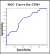
Methods: The sample was composed of 43 archived specimens. Clinical features and histological grade of malignancy were obtained. The infiltrating intensity of CD4 +, CD8 positive cells were assessed by immunohistochemistry. One-way ANOVA was used to study the association between CD4 +, CD8 + and the grade of OSCC. The cut-off values of the proposed diagnostic indices were received from calculating the coordinates of the receiver operating characteristic (ROC) curve. For clinicopathologic data Independent-Samples T test, Pearson Correlation Coefficient, Correlation Coefficient were used clinicopathologic characteristics.
Results: CD4 +and CD8 + were observed in all specimens. CD4 + were more frequent in poorly differentiated specimens (74.14) (P= 0.021<0.05). CD8 + were more frequent in well- differentiated specimens (51.18). None of these correlations were significant (P=0.454>0.05). CD4 +/ CD8 ratio was higher in low-grade specimens (180.28) (P=0.017<0.05). No differences between CD4 +, CD8 +and CD4 +/ CD8 ratio between poorly- differentiated and moderately- differentiated groups ROC P value (0.370, 0.248, 0.126) respectively. there is a difference between CD4 +, CD4 +/ CD8ratio between poorly- differentiated and well- differentiated groups ROC P value (0.022, 0.341, 0.012) Sensitivity (0.857, 0.882), specificity (0.706, 0.857) respectively. and no differences between CD8 + poorly- differentiated and well- differentiated groups ROC P value (0.341). there is a difference between CD4 + between moderately - differentiated and well- differentiated groups ROC P value (0.038) Sensitivity (0.368), specificity (0,765). No significant correlation was obtained with clinicopathologic findings of OSCC.
Conclusion: CD4 + and CD4 +/ CD8 + ratio are independent prognostic factor in OSCC.
Keywords: CD4 +; CD8 +; OSCC; Prognosis; TILs.
Conflict of interest statement
The authors declare that they have no conflict of interest.
Figures


References
-
- Bloebaum M, Poort L, Böckmann R, et al. Survival after curative surgical treatment for primary oral squamous cell carcinoma. J Craniomaxillofac Surg. 2014;42:1572–6. - PubMed
-
- Cho YA, Yoon HJ, Lee JI, et al. Relationship between the expressions of PD-L1 and tumor-infiltrating lymphocytes in oral squamous cell carcinoma. Oral Oncol. 2011;47:1148–53. - PubMed
MeSH terms
LinkOut - more resources
Full Text Sources
Medical
Research Materials

