. 2023 Feb:59:102584.
doi: 10.1016/j.redox.2022.102584. Epub 2022 Dec 22.

Ferroptosis is critical for phthalates driving the blood-testis barrier dysfunction via targeting transferrin receptor

Affiliations

Ferroptosis is critical for phthalates driving the blood-testis barrier dysfunction via targeting transferrin receptor

Yi Zhao et al. Redox Biol. 2023 Feb.

Abstract

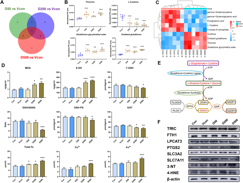
The global rate of human male infertility is rising at an alarming rate owing to environmental and lifestyle changes. Phthalates are the most hazardous chemical additives in plastics and have an apparently negative impact on the function of male reproductive system. Ferroptosis is a recently described form of iron-dependent cell death and has been linked to several diseases. Transferrin receptor (TfRC), a specific ferroptosis marker, is a universal iron importer for all cells using extracellular transferrin. We aim to investigate the potential involvement of ferroptosis during male reproductive toxicity, and provide means for drawing conclusions on the effect of ferroptosis in phthalates-induced male reproductive disease. In this study, we found that di (2-ethylhexyl) phthalate (DEHP) triggered blood-testis barrier (BTB) dysfunction in the mouse testicular tissues. DEHP also induced mitochondrial morphological changes and lipid peroxidation, which are manifestations of ferroptosis. As the primary metabolite of DEHP, mono-2-ethylhexyl phthalate (MEHP) induced ferroptosis by inhibiting glutathione defense network and increasing lipid peroxidation. TfRC knockdown blocked MEHP-induced ferroptosis by decreasing mitochondrial and intracellular levels of Fe2+. Our findings indicate that TfRC can regulate Sertoli cell ferroptosis and therefore is a novel therapeutic molecule for reproductive disorders in male patients with infertility.

Keywords: Blood-testis barrier; Di (2-ethylhexyl) phthalate; Ferroptosis; Sertoli cells; Transferrin receptor.


Conflict of interest statement

Declaration of competing interest The authors declare that they have no conflict of interest.

Figures

Image 1
Graphical abstract
Fig. 1
Fig. 1
DEHP induces structural and functional damage to Sertoli cells. (A) The mice were treated with water, corn oil and DEHP at a dose of 50, 200, and 500 mg/kg/d (D50, D200, and D500, respectively) by oral gavage for 28 days. (B) Testis coefficient. (C) Histopathology with H&E, staining. (D) Johnsen Score. (E) AR, ABP, FSHR and Tf protein levels in mice testes; β-actin served as loading control for the total fraction. (F) Changes of BTB structure in mice. (G) The function and integrity BTB in mouse testis. (H) BTB-related protein levels in mice testes; β-actin served as loading control for the total fraction. (I) Transmission electron microscopy and Flameng scores; yellow: mitochondria. (J) Eosin staining of mouse sperm, Papanicolaou staining of mouse sperm and sperm malformation. Data are presented as the mean ± SD (n = 3). Symbol for the significance of differences between the Vcon group and another group: **P < 0.01, ***P < 0.001. (For interpretation of the references to colour in this figure legend, the reader is referred to the Web version of this article.)
Fig. 2
Fig. 2
DEHP promotes glutathione metabolism disorders and induces ferroptosis in mouse testes. (A) Venn diagram of metabolite profiling in the testes of mice exposed to D50, D200 and D500. (B) Thiourea, l-Cysteine, glutathione glycylmenthyl ester and oxidized glutathione are the three most significantly differential metabolites. (C) Heat map of metabolite profiling in testes of mice exposed to D50, D200 and D500. (D) T-GSH contents, the GSH/GSSG ratio, GSH-PX activity, GST activity, MDA content, 8-OH content, and total Fe, Fe2+ and Fe3+ contents of mouse testes. (E) Schematic diagram depicting the regulation of DEHP induced glutathione metabolism dysfunction in the testes of mice. (F) Levels of ferroptosis-related proteins in mouse testes; β-actin served as loading controls for the total fraction. Data are presented as the mean ± SD (n = 3). Symbol for the significance of differences between the Vcon group and another group: *P < 0.05, **P < 0.01, ***P < 0.001.
Fig. 3
Fig. 3
MEHP induces structural and functional damage to TM4 cells. (A) TM4 cells were treated with MEHP. (B) The cell viability was assayed using CCK-8 after exposure to different concentrations of MEHP in TM4 cells. (C) Transmission electron microscopy analysis; yellow indicates mitochondria. (D) Determination of mitochondrial average surface area. (E) Flameng scores. (F) Heat map of relative mRNA levels of secretory dysfunction in TM4 cells. (G) Levels of AR, ABP, FSHR and Tf in TM4 cells; β-actin served as loading controls for the total fraction. (H) Quantitation of the protein expression of AR, ABP, FSHR and Tf in TM4 cells. (I) GSEA analysis revealed a decreased cell junction assembly signaling pathway. (J) GSEA analysis revealed a decreased cell junction organization assembly signaling pathway. (K) Changes of TER in SCs. Data are presented as the mean ± SD (n = 3). Symbol for the significance of differences between the DMSO group and another group: *P < 0.05, **P < 0.01, ***P < 0.001. (For interpretation of the references to colour in this figure legend, the reader is referred to the Web version of this article.)
Fig. 4
Fig. 4
MEHP promotes glutathione metabolism disorders and induces ferroptosis in TM4 cells. (A) GO analysis based on RNA-sequencing results of TM4 cells after exposure to M400. (B) BP analysis based on RNA-sequencing results of TM4 cells after exposure to M400. (C) KEGG analysis based on RNA-sequencing results of TM4 cells after exposure to M400. (D) Heat map of relative mRNA levels of glutathione metabolism-related genes in M400-treated TM4 cells. (E) T-GSH contents, the GSH/GSSG ratio, GSH-PX activity, LPO contents, MDA contents, 8-OH contents in TM4 cells. (F) GSEA analysis revealed an increased ferroptosis assembly signaling pathway. (G) Levels of ferroptosis-related proteins in TM4 cells after exposure to M400; β-actin served as loading controls for the total fraction. (H) Lipid ROS, mitochondrial ROS, mitochondrial iron and intracellular iron level in TM4 cells. Data are presented as the mean ± SD (n = 3). Symbol for the significance of differences between the DMSO group and another group: *P < 0.05, **P < 0.01, ***P < 0.001.
Fig. 5
Fig. 5
Fer-1 inhibits MEHP- induced ferroptosis in TM4 cells. (A) TM4 cells were treated with Fer-1 and DFO, Erastin, or M400; their cell viability was assayed using a CCK8 assay (B) Transmission electron microscopy analysis, mitochondrial average surface area measurement, and Flameng scorea of the treated TM4 cells; yellow indicates mitochondria; green indicates autophagic vacuole. (C) T-GSH contents, the GSH/GSSG ratio, GSH-PX activity, LPO contents, MDA contents, and 8-OH content of TM4 cells. (D) Intracellular and lipid ROS production of TM4 cells. (E) Mitochondrial ROS production of TM4 cells. Data are presented as the mean ± SD (n = 3). Symbol for the significance of differences between the DMSO group and another group: *P < 0.05, **P < 0.01, ***P < 0.001. Symbol for the significance of differences between the MEHP groups and Fer-1 + M400 group groups: ##P < 0.01, ###P < 0.001. (For interpretation of the references to colour in this figure legend, the reader is referred to the Web version of this article.)
Fig. 6
Fig. 6
Fer-1 and DFO inhibit MEHP-induced change in iron levels in TM4 cells. (A) Expression of ferroptosis-related proteins in TM4 cells, β-actin served as loading controls for the total fraction. (B) Quantitation of the protein expression of ferroptosis-related proteins in TM4 cells. (C) Intracellular iron level of TM4 cells. (D) Intracellular iron level in TM4 cells. Data are presented as the mean ± SD (n = 3). Symbol for the significance of differences between the DMSO group and another group: *P < 0.05, **P < 0.01, ***P < 0.001. Symbol for the significance of differences between the MEHP groups and Fer-1/DFO + M400 group groups: #P < 0.05, ###P < 0.001.
Fig. 7
Fig. 7
TfRC knockdown inhibits MEHP-induced ferroptosis in TM4 cells. (A) TM4 cells were treated with siTfRC and/or M400. (B) The cell viability was assayed using a CCK8 assay. (C) Levels of TfRC in TM4 cells. (D) Quantitation of the protein expression of TfRC. (E) Intracellular and lipid ROS generation; mitochondrial ROS; intracellular iron level; mitochondrial iron level. (F) T-GSH contents, the GSH/GSSG ratio, GSH-PX activity, LPO contents, MDA contents, and 8-OH content, T-AOC activity, T-SOD activity and CAT activity. (G) Changes of TER in SCs. (H) Molecular docking simulation for the ligand–protein binding of MEHP with TfRC. (I) Schematic diagram depicting the regulation of MEHP-induced ferroptosis by increasing TfRC level in Sertoli cells. Data are presented as the mean ± SD (n = 3). Symbol for the significance of differences between the DMSO group and another group: *P < 0.05, **P < 0.01, ***P < 0.001. Symbol for the significance of differences between the siNC + M400 group and siTf + M400 group: #P < 0.05, ##P < 0.01, ###P < 0.001.
Fig. 8
Fig. 8
DEHP promotes ferroptosis by targeting TfRC in Sertoli cells, thereby inducing BTB dysfunction.

References

    1. Ferguson K.K., McElrath T.F., Meeker J.D. Environmental phthalate exposure and preterm birth. JAMA Pediatr. 2014;168:61–67. - PMC - PubMed
    1. Diamanti-Kandarakis E., Bourguignon J.P., Giudice L.C., Hauser R., Prins G.S., Soto A.M., Zoeller R.T., Gore A.C. Endocrine-disrupting chemicals: an Endocrine Society scientific statement. Endocr. Rev. 2009;30:293–342. - PMC - PubMed
    1. Zhou Y., Wang H., Chen Y., Jiang Q. Environmental and food contamination with plasticisers in China. Lancet. 2011;378:e4. - PubMed
    1. IHS, Plasticizers 2021. https://ihsmarkit.com/products/plasticizers-chemical-economics-handbook.... Online Published May.
    1. Dai Y.X., Zhu S.Y., Chen J., Li M.Z., Talukder M., Li J.L. Role of Toll-like Receptor/MyD88 Signaling in Lycopene Alleviated Di-2-ethylhexyl Phthalate (DEHP)-Induced Inflammatory Response. J. Agric. Food Chem. 2022;70:10022–10030. - PubMed

Publication types