The Location of the Parasympathetic Fibres within the Vagus Nerve Rootlets: A Case Report and a Review of the Literature
- PMID: 36580909
- PMCID: PMC9986834
- DOI: 10.1159/000528094
The Location of the Parasympathetic Fibres within the Vagus Nerve Rootlets: A Case Report and a Review of the Literature
Abstract
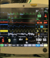
The vagus nerve has motor, sensory, and parasympathetic components. Understanding the nerve's internal anatomy, its variations, and relationship to the glossopharyngeal nerve are crucial for neurosurgeons decompressing the lower cranial nerves. We present a case report demonstrating the location of the parasympathetic fibres within the vagus nerve rootlets. A 47-year-old woman presented with a 1-year history of medically refractory left-sided glossopharyngeal neuralgia and a more recent history of left-sided hemi-laryngopharyngeal spasm. magnetic resonance imaging showed her left posterior inferior cerebellar artery distorting the lower cranial nerves on the affected left side. The patient consented to microvascular decompression of the lower cranial nerves with possible sectioning of the glossopharyngeal and upper sensory rootlets of the vagus nerve. During surgery, electrical stimulation of the most caudal rootlet of the vagus nerve triggered profound bradycardia. None of the more rostral rootlets had a similar parasympathetic response. This case is the first demonstration, to our knowledge, of the location of the cardiac parasympathetic fibres within the human vagus nerve rootlets. This new understanding of the vagus nerve rootlets' distribution of pure sensory (most rostral), motor/sensory (more caudal), and parasympathetic (most caudal) fibres may lead to a better understanding and diagnosis of the vagal rhizopathies. Approximately 20% of patients with glossopharyngeal neuralgia also have paroxysmal cough. This could be due to the anatomical juxtaposition of the IXth cranial nerve with the rostral vagal rootlets with pure sensory fibres (which mediate a tickling sensation in the lungs). A subgroup of patients with glossopharyngeal neuralgia have neuralgia-induced syncope. The cause of this rare condition, "vago-glossopharyngeal neuralgia," has been debated since it was first described by Riley in 1942. Our case supports the theory that this neuralgia-induced bradycardia is reflexively mediated through the brainstem with afferent impulses in the IXth and efferent impulses in the Xth cranial nerve. The rarer co-occurrence of glossopharyngeal neuralgia with hemi-laryngopharyngeal spasm (as seen in this case) may be explained by the proximity of the IXth nerve with the more caudal vagus rootlets which have motor (and probably sensory) supply to the throat. Finally, if there is a vagal rhizopathy related to compression of its parasympathetic fibres, one would expect it to be at the most caudal rootlet of the vagus nerve.
Keywords: Glossopharyngeal nerve; Glossopharyngeal neuralgia; Microvascular decompression; Parasympathetic vagus nerve; Vago-glossopharyngeal neuralgia.
© 2022 The Author(s). Published by S. Karger AG, Basel.
Conflict of interest statement
The authors have no personal or institutional interest to declare.
Figures


References
-
- Yuan H, Silberstein SD. Vagus nerve and vagus nerve stimulation a comprehensive review Part I. Headache. 2016 Jan;56((1)):71–78. - PubMed
-
- Kenny BJ, Bordoni B. Neuroanatomy cranial nerve 10 (vagus nerve) StatPearls Publishing. 2021 Nov 14; InStatPearls [Internet] - PubMed
-
- Honey CR, Gooderham P, Morrison M, Ivanishvili Z. Episodic hemilaryngopharyngeal spasm (HELPS) syndrome case report of a surgically treatable novel neuropathy. J Neurosurg. 2017 May 1;126((5)):1653–1656. - PubMed
-
- Honey CR, Krüger MT, Morrison MD, Dhaliwal BS, Hu A. Vagus associated neurogenic cough occurring due to unilateral vascular encroachment of its Root a case report and proof of concept of VANCOUVER syndrome. Ann Otol Rhinol Laryngol. 2020 May;129((5)):523–527. - PubMed
-
- Harries AM, Dong CCJ, Honey CR. Use of endotracheal tube electrodes in treating glossopharyngeal neuralgia technical note. Stereotact Funct Neurosurg. 2012;90((3)):141–144. - PubMed
Publication types
MeSH terms
LinkOut - more resources
Full Text Sources

