Review
. 2023 Feb;299(2):102838.
doi: 10.1016/j.jbc.2022.102838. Epub 2022 Dec 26.

Regulation and function of the mammalian tricarboxylic acid cycle

Affiliations
Review

Regulation and function of the mammalian tricarboxylic acid cycle

Paige K Arnold et al. J Biol Chem. 2023 Feb.

Abstract

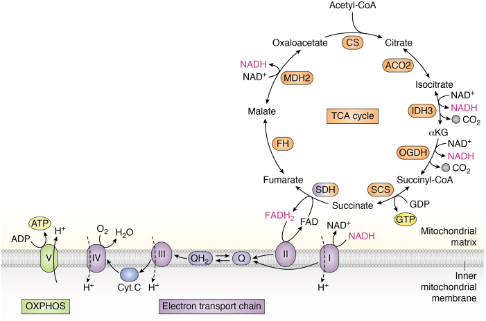
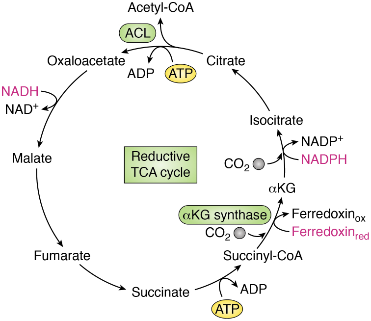
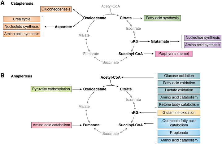
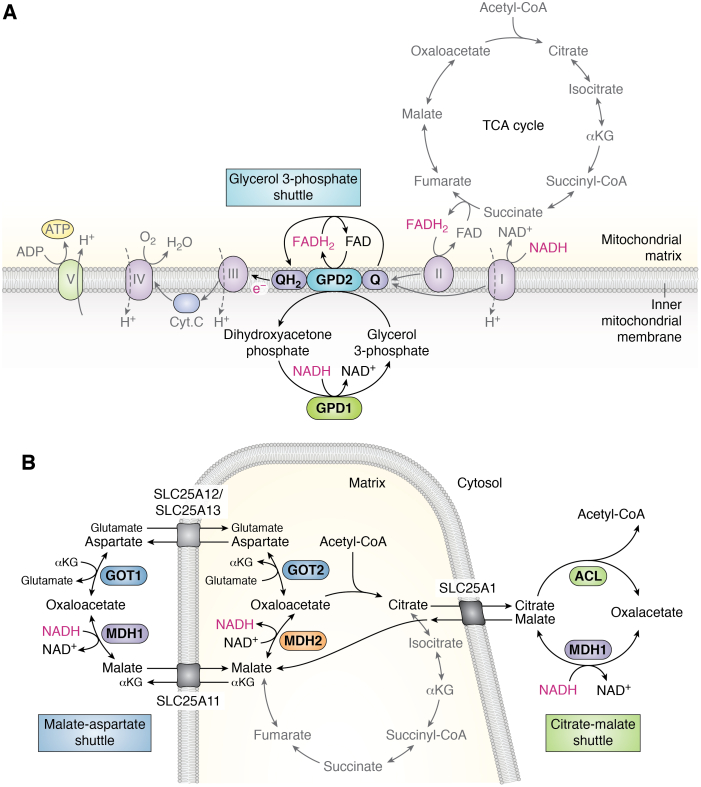
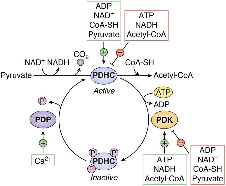
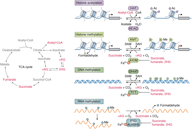
The tricarboxylic acid (TCA) cycle, otherwise known as the Krebs cycle, is a central metabolic pathway that performs the essential function of oxidizing nutrients to support cellular bioenergetics. More recently, it has become evident that TCA cycle behavior is dynamic, and products of the TCA cycle can be co-opted in cancer and other pathologic states. In this review, we revisit the TCA cycle, including its potential origins and the history of its discovery. We provide a detailed accounting of the requirements for sustained TCA cycle function and the critical regulatory nodes that can stimulate or constrain TCA cycle activity. We also discuss recent advances in our understanding of the flexibility of TCA cycle wiring and the increasingly appreciated heterogeneity in TCA cycle activity exhibited by mammalian cells. Deeper insight into how the TCA cycle can be differentially regulated and, consequently, configured in different contexts will shed light on how this pathway is primed to meet the requirements of distinct mammalian cell states.

Keywords: Krebs cycle; bioenergetics; cell metabolism; citric acid cycle; tricarboxylic acid cycle.


Conflict of interest statement

Conflict of interest P. K. A. and L. W. S. F. are authors on patent applications that cover links between cellular metabolism and cell fate control.

Figures

Figure 1
Figure 1
Overview of the tricarboxylic acid (TCA) cycle and electron transport chain (ETC). The TCA cycle starts when the two-carbon molecule acetyl-CoA combines with four-carbon oxaloacetate to form citrate, a reaction catalyzed by citrate synthase (CS). Citrate is then converted to isocitrate by aconitase 2 (ACO2). Isocitrate is decarboxylated to alpha-ketoglutarate (αKG) in an NAD+-dependent manner by isocitrate dehydrogenase 3 (IDH3) or in an NADP+-dependent manner by isocitrate dehydrogenase 2 (IDH2), releasing carbon dioxide (CO2). αKG undergoes decarboxylation to succinyl-CoA via the oxoglutarate dehydrogenase complex (OGDH), producing NADH and releasing CO2. Succinyl-CoA is then converted to succinate by succinyl-CoA synthetase (SCS). This is the only substrate-level phosphorylation step in the TCA cycle, as it is coupled to the generation of GTP or ATP. Succinate is converted to fumarate by succinate dehydrogenase (SDH) complex, a multisubunit enzyme complex that participates in both the TCA cycle and the electron transport chain (ETC). SDH reduces FAD to FADH2, which donates its electrons to complex II. Fumarate is converted to malate by fumarate hydratase (FH). Malate dehydrogenase 2 (MDH2) converts malate to oxaloacetate in an NAD+-dependent manner, regenerating the starting molecule and supporting the next turn of the cycle. Note: most TCA cycle reactions are reversible. Substrate oxidation reactions are coupled to reduction of electron carriers NAD+ and FAD: each complete turn of the TCA cycle generates three NADH and one FADH2 molecules, which donate their electrons to complex I and complex II, respectively. These reducing equivalents are reoxidized upon donating their electrons to the ETC, supporting continued TCA cycle activity. Electrons donated to complexes I and II are transferred to ubiquinone (Q), reducing it to ubiquinol (QH2). Ubiquinol is reoxidized to ubiquinone upon passing its electrons to complex III, which transfers electrons to cytochrome C (Cyt C). Cyt C passes its electrons onto complex IV, which then transfers its electrons to the terminal electron acceptor, oxygen (O2), forming water (H2O). As electrons are transferred through the ETC and eventually onto oxygen, complexes I, III, and IV pump protons across the inner mitochondrial membrane. This proton pumping establishes a proton gradient that is used by complex V, or ATP synthase, to generate ATP from ADP, a process known as oxidative phosphorylation (OXPHOS). TCA cycle enzymes are colored in orange; SDH is colored blue and orange because it participates in both the TCA cycle and the ETC. Reducing equivalents are shown in pink.
Figure 2
Figure 2
The reductive tricarboxylic acid (TCA) cycle. A simplified schematic depicting the reductive TCA cycle or reverse Krebs cycle. Most reactions of this cycle are the same as those of the oxidative TCA cycle but in reverse and are catalyzed by similar enzymes. The major exceptions include (1) cleavage of citrate to form oxaloacetate and acetyl-CoA and (2) the production of alpha-ketoglutarate (αKG) from succinyl-CoA. Citrate cleavage requires ATP and is carried out by ATP-citrate lyase (ACL) or the related citryl-CoA lyase and citryl-CoA synthase enzymes. Conversion of succinyl-CoA to αKG, mediated by αKG synthase, is highly energetically unfavorable and thus requires a strong reducing agent in the form of reduced ferredoxin (Ferredoxinred). While the oxidative TCA cycle combusts carbon and produces reducing equivalents that drive ATP synthesis, the reductive TCA cycle consumes ATP and reducing equivalents to assimilate carbon and produce acetyl-CoA. Reducing equivalents are shown in pink.
Figure 3
Figure 3
Outputs and inputs into the tricarboxylic acid (TCA) cycle. A functioning TCA cycle requires a continuous pool of acetyl-CoA and supply of TCA cycle intermediates that can be used to synthesize oxaloacetate. A, removal of TCA cycle intermediates (“cataplerosis”) occurs at multiple steps of the cycle to supply precursors for biosynthetic processes or feed into other metabolic pathways. B, replacement of TCA cycle intermediates (“anaplerosis”) is required to support continuous production of oxaloacetate. Sources of anaplerosis are shown in this panel. Several pathways either produce acetyl-CoA directly or produce pyruvate, an indirect source of acetyl-CoA through the activity of the pyruvate dehydrogenase complex.
Figure 4
Figure 4
Electron shuttles that intersect with the tricarboxylic acid (TCA) cycle or electron transport chain (ETC).A, in the glycerol 3-phosphate shuttle, the conversion of dihydroxyacetone phosphate into glycerol 3-phosphate by cytosolic glycerol 3-phosphate dehydrogenase 1 (GPD1) regenerates cytosolic NAD+ in the cytoplasm to support continued glycolysis. Glycerol 3-phosphate is subsequently converted back to dihydroxyacetone phosphate on the outer side of the inner mitochondrial membrane by mitochondrial glycerol 3-phosphate dehydrogenase 2 (GPD2), which is coupled with the conversion of FAD to FADH2. FADH2 donates electrons to ubiquinone (Q), reducing it to ubiquinol (QH2) that passes its electrons to complex III of the ETC. B, in the malate–aspartate shuttle (left), the TCA cycle intermediate oxaloacetate (OAA) and glutamate undergo transamination by glutamic-oxaloacetic transaminase 2 (GOT2), producing alpha-ketoglutarate (αKG) and aspartate. Mitochondrial aspartate is exported to the cytoplasm by the mitochondrial transporter proteins SLC25A12 or SLC25A13, and this efflux is concomitant with import of glutamate and a proton. Cytosolic aspartate is consumed by the cytosolic transaminase GOT1, converting αKG to glutamate and producing OAA. Cytosolic OAA is converted to malate by malate dehydrogenase 1 (MDH1), which is coupled with oxidation of NADH to NAD+, supporting glycolysis by regenerating NAD+ required for GAPDH. MDH1-generated malate is then reimported into mitochondria via the transporter SLC25A11, which is coupled with efflux of mitochondrial αKG. MDH2 converts malate to OAA, thereby reducing NAD+ to NADH and completing the cycle. In the citrate–malate shuttle (right), the citrate–malate antiporter SLC25A1 exports citrate to the cytosol, where it undergoes energy-dependent cleavage by ATP-citrate lyase (ACL), liberating acetyl-CoA and OAA. Conversion of OAA to malate by MDH1 supports NADH oxidation to NAD+, and this malate is imported into mitochondria in exchange for citrate by SLC25A1. In the mitochondria, malate is oxidized to OAA by MDH2, completing the shuttling of NADH into the mitochondrion.
Figure 5
Figure 5
Allosteric and covalent regulation of the pyruvate dehydrogenase (PDHC) complex. The PDHC catalyzes the irreversible decarboxylation of pyruvate to acetyl-CoA, releasing CO2 and transferring electrons to NAD+ to form NADH. The PDHC utilizes multiple coenzymes, including coenzyme A (CoA-SH), during its multistep reaction. The complex is activated by increased levels of ADP, NAD+, CoA-SH, and its substrate pyruvate. Conversely, the PDHC is allosterically inhibited by high levels of ATP, NADH, and its product acetyl-CoA. Phosphorylation of any of three serine residues on the PDHC by pyruvate dehydrogenase kinases (PDKs) inactivates the complex. The PDKs are activated by high levels of ATP, NADH, and acetyl-CoA, reinforcing the shutdown of PDHC flux under these high-energy conditions. Reciprocally, the PDKs are inhibited by ADP, NAD+, CoA-SH, and pyruvate. The inhibitory phosphorylation of PDHC is reversible and can be removed by the pyruvate dehydrogenase phosphatases (PDPs), which are activated by mitochondrial calcium (Ca2+).
Figure 6
Figure 6
Regulation of chromatin by tricarboxylic acid (TCA) cycle–associated metabolites. Certain TCA cycle–derived metabolites (left) control the regulation of chromatin and, in turn, gene expression. Histone acetyltransferases (HAT) transfer acetyl groups from acetyl-CoA to histones, thereby altering chromatin accessibility. Histone deacetylases (HDACs) remove acetyl groups from histones and generate acetate as a product. Methylation on histones and DNA is deposited by histone lysine methyltransferases (KMTs) and DNA methyltransferases (DNMTs), respectively. DNMT and KMT add methyl groups by catalyzing the transfer of a methyl group from S-adenosyl methionine (SAM), producing S-adenosyl homocysteine (SAH). Alpha-ketoglutarate (αKG)-dependent dioxygenases regulate the demethylation of histones and nucleic acids. Jumonji C-domain–containing histone demethylases (JHDMs) and ten–eleven translocation (TET) DNA methylcytosine dioxygenases, which remove repressive histone marks and 5-methylcytosine, respectively, require αKG as an obligate cosubstrate and are competitively inhibited by succinate, fumarate, and 2-hydroxyglutarate (2HG). mRNA methylation is controlled by the RNA methyltransferases and αKG-dependent dioxygenases FTO and ALKBH5, which like all αKG-dependent dioxygenases are stimulated by αKG and repressed by succinate, fumarate, and 2HG. Histone and RNA demethylation produce formaldehyde as a byproduct.

References

    1. Krebs H.A., Johnson W.A. Metabolism of ketonic acids in animal tissues. Biochem. J. 1937;31:645–660. - PMC - PubMed
    1. Krebs H.A., Johnson W.A. The role of citric acid in intermediate metabolism in animal tissues. FEBS Lett. 1980;117 doi: 10.4159/harvard.9780674366701.c143. - DOI - PubMed
    1. Ryan D.G., O'Neill L.A.J. Krebs cycle Reborn in macrophage immunometabolism. Annu. Rev. Immunol. 2020;38:289–313. - PubMed
    1. Krebs H.A., Eggleston L.V. The oxidation of pyruvate in pigeon breast muscle. Biochem. J. 1940;34:442–459. - PMC - PubMed
    1. Inigo M., Deja S., Burgess S.C. Ins and outs of the TCA cycle: the central role of anaplerosis. Annu. Rev. Nutr. 2021;41:19–47. - PubMed

Publication types

LinkOut - more resources