. 2022 Dec 29;12(1):22553.
doi: 10.1038/s41598-022-27082-1.

Implementation of a three-dimensional (3D) robotic digital microscope (AEOS) in spinal procedures

Affiliations

Implementation of a three-dimensional (3D) robotic digital microscope (AEOS) in spinal procedures

Stefan Motov et al. Sci Rep. .

Abstract

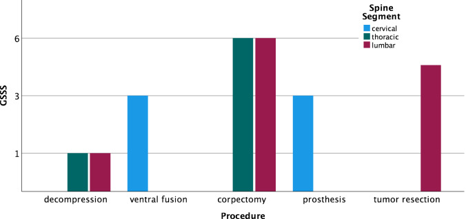
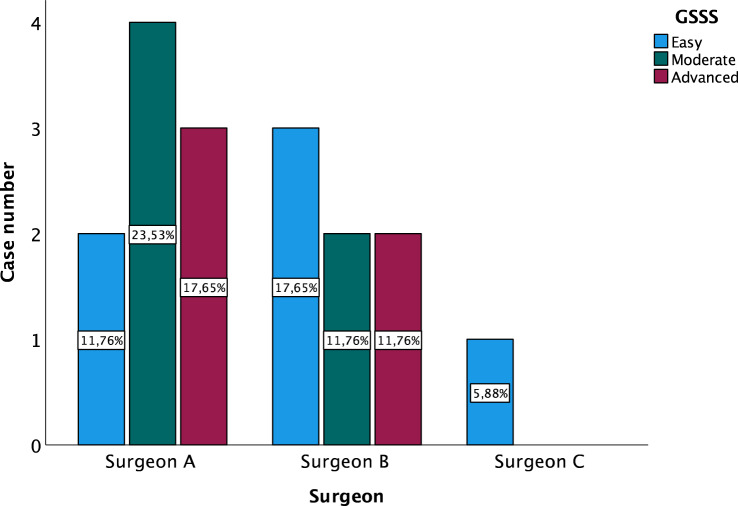
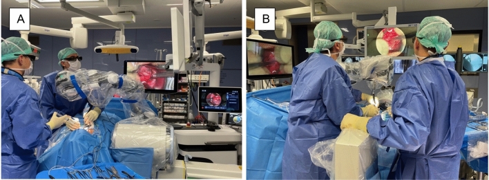
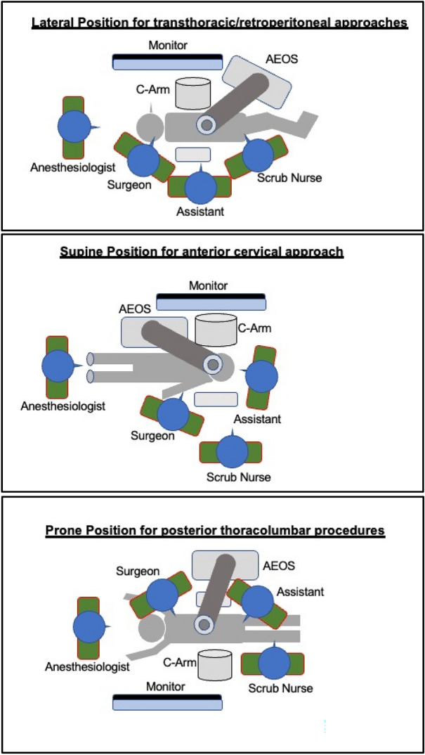
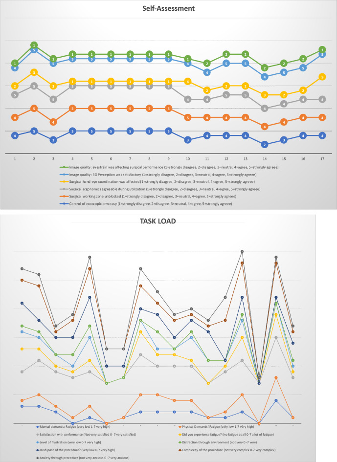
Three-dimensional exoscopes have been designed to overcome certain insufficiencies of operative microscopes. We aimed to explore the clinical use in various spinal surgeries. We performed surgery on patients with different spine entities in a neurosurgical department according to the current standard operating procedures over a 4-week period of time. The microsurgical part has been performed with Aesculap AEOS 3D microscope. Three neurosurgeons with different degree of surgical expertise completed a questionnaire with 43 items based on intraoperative handling and feasibility after the procedures. We collected and analyzed data from seventeen patients (35% male/65% female) with a median age of 70 years [CI 47-86] and median BMI of 25.8 kg/m2 [range 21-33]. We included a variety of spinal pathologies (10 degenerative, 4 tumor and 3 infectious cases) with different level of complexity. Regarding setup conflicts we observed issues with adjustment of the monitor position or while using additional equipment (e.g. fluoroscopy in fusion surgery) (p = 0.007/p = 0.001). However image resolution and sharpness as well as 3D-depth perception were completely satisfactory for all surgeons in all procedures. The utilization of the exoscopic arm was easy for 76.5% of the surgeons, and all of them declared a significant improvement of the surgical corridor. The 3D-exoscope implementation appears to achieve very satisfactory results in spinal procedures especially with minimally invasive approaches.


Conflict of interest statement

The authors declare no competing interests.

Figures

Figure 1
Figure 1
Case complexity based on spine segment and GSSS.
Figure 2
Figure 2
Case complexity distribution between surgeons based on GSSS.
Figure 3
Figure 3
Blood loss distribution based on BMI and surgeon.
Figure 4
Figure 4
Surgery times (minutes) based on BMI and surgeon.
Figure 5
Figure 5
OR setup for ACDF: Surgical corridor remains unblocked with a good visualization for both surgeon and assistant. (A) Assistant‘s and OR nurse‘s 55 inch monitor is directly opposite of them; (B) 32 inch monitor is in front of the surgeon, fluoroscope is placed in lateral position
Figure 6
Figure 6
OR setup for cervical and thoracolumbar procedures based on patient and equipment positioning.
Figure 7
Figure 7
Exemplary case 1: anterior cervical discectomy and disc replacement or fusion. (A) Anterior discectomy and removal of the anterior longitudinal ligament; (B) Cervical disc replacement with prosthesis; (C) Cervical foraminotomy; (D) Saving two different targets of focus enables semi-automatic adjustment between the two levels of interest
Figure 8
Figure 8
Exemplary case 2: thoracic corpectomy and vertebral body replacement with a carbon fiber-reinforced PEEK implant for metastatic disease. (A) Transpleural minithoracotomy approach for vertebral body resection; (B) Vizualization of vertebral body replacement; (C) Providing different angles of view in the depth enables adequate preparation and hemostasis; (D) Watertight closure of pleural cavity after chest tube placement
Figure 9
Figure 9
Results of surgeons’ questionnaires classified in the categories self-assessment, task load and utilization and image quality.
Figure 9
Figure 9
Results of surgeons’ questionnaires classified in the categories self-assessment, task load and utilization and image quality.

References

    1. Amoo M, Henry J, Javadpour M. Beyond magnification and illumination: Preliminary clinical experience with the 4K 3D ORBEYE exoscope and a literature review. Acta Neurochir. (Wien) 2021;163(8):2107–2115. doi: 10.1007/s00701-021-04838-8. - DOI - PubMed
    1. Siller S, et al. A high-definition 3D exoscope as an alternative to the operating microscope in spinal microsurgery. J. Neurosurg. Spine. 2020 doi: 10.3171/2020.4.SPINE20374. - DOI - PubMed
    1. Maurer S, et al. Evaluation of a novel three-dimensional robotic digital microscope (aeos) in neurosurgery. Cancers (Basel) 2021 doi: 10.3390/cancers13174273. - DOI - PMC - PubMed
    1. Muhammad S, Lehecka M, Niemela M. Preliminary experience with a digital robotic exoscope in cranial and spinal surgery: A review of the Synaptive Modus V system. Acta Neurochir. (Wien) 2019;161(10):2175–2180. doi: 10.1007/s00701-019-03953-x. - DOI - PMC - PubMed
    1. Montemurro N, et al. The exoscope in neurosurgery: An overview of the current literature of intraoperative use in brain and spine surgery. J. Clin. Med. 2021 doi: 10.3390/jcm11010223. - DOI - PMC - PubMed

Publication types