. 2022 Dec 29;21(1):228.
doi: 10.1186/s12943-022-01697-4.

Targeting SOST using a small-molecule compound retards breast cancer bone metastasis

Affiliations

Targeting SOST using a small-molecule compound retards breast cancer bone metastasis

Lisha Sun et al. Mol Cancer. .

Erratum in

Abstract

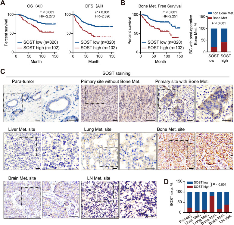
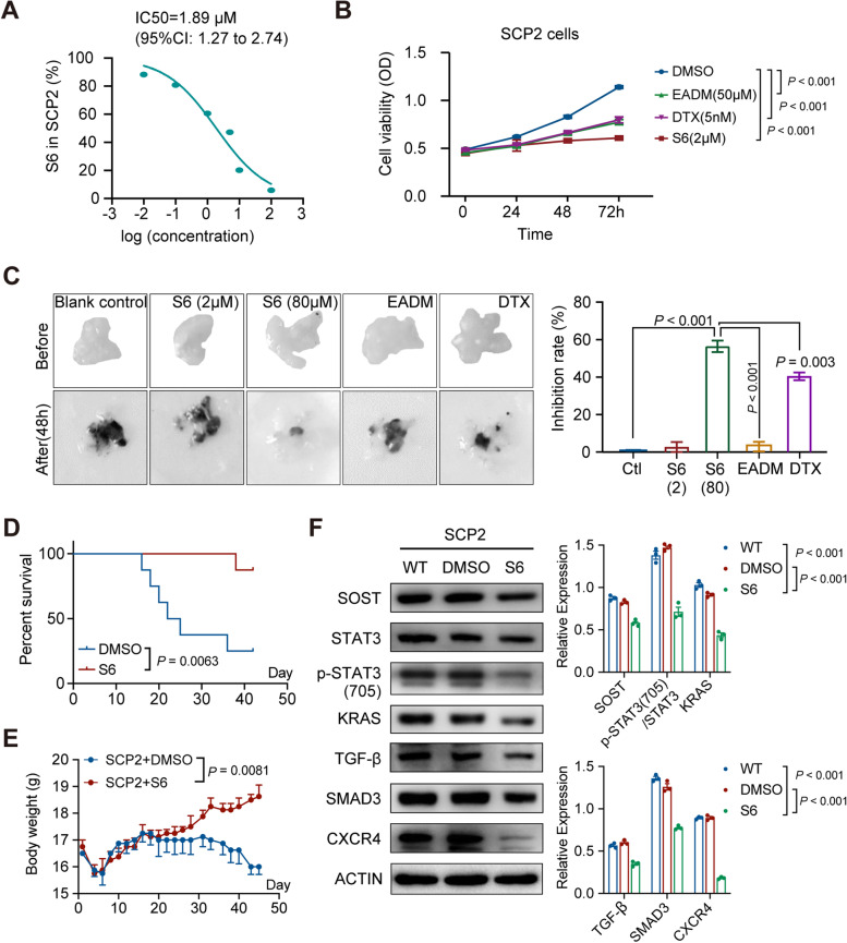
Background: Breast cancer metastasis to the bone can be exacerbated by osteoporosis, is associated with poor long-term survival, and has limited therapeutic options. Sclerostin (SOST) is an endogenous inhibitor of bone formation, and an attractive target for treatment of osteoporosis. However, it is unclear whether SOST can be used as a therapeutic target for bone metastases of breast cancer, and whether small molecule compounds that target SOST in breast cancer cells can inhibit breast cancer bone metastasis.

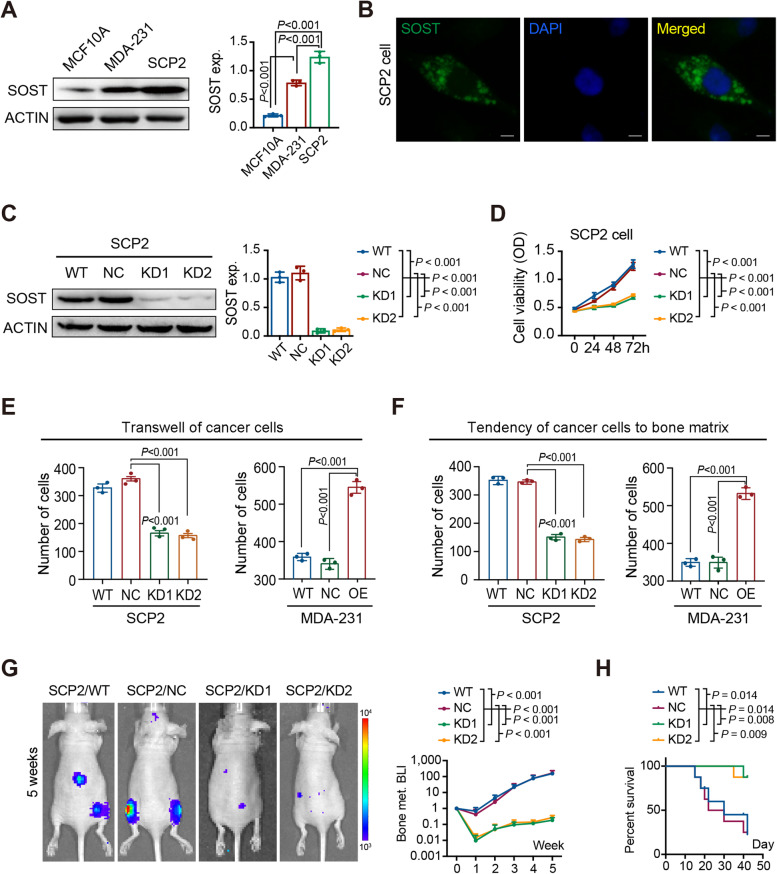
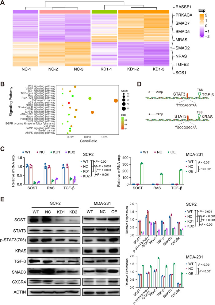
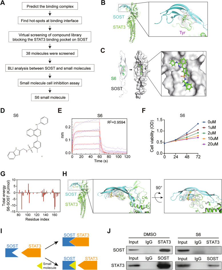
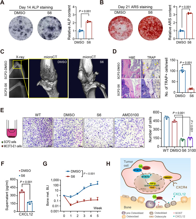
Methods: SOST expression in 442 breast cancer tissues was characterized by immunohistochemistry and statistically analyzed for the association with breast cancer bone metastases. Bone metastatic breast cancer SCP2 cells were induced for SOST silencing or overexpression and their bone metastatic behaviors were tested in vitro and in vivo. To identify potential therapeutics, we screened inhibitors of the interaction of SOST with STAT3 from a small chemical molecule library and tested the inhibitory effects of one inhibitor on breast cancer growth and bone metastasis in vitro and in vivo.

Results: We found that up-regulated SOST expression was associated with breast cancer bone metastases and worse survival of breast cancer patients. SOST silencing significantly reduced the bone metastatic capacity of SCP2 cells. SOST interacted with STAT3 to enhance the TGF-β/KRAS signaling, increasing both tumor growth and bone metastasis. Treatment with one lead candidate, S6, significantly inhibited the growth of breast-cancer organoids and bone metastasis in mice.

Conclusions: Our findings highlight a new class of potential therapeutics for treatment of bone metastasis in breast cancer.

Keywords: Bone metastasis; Breast cancer; SOST; Small-molecule compound.


Conflict of interest statement

The authors declare that they have no competing interests.

Figures

Fig. 1
Fig. 1
Up-regulated SOST expression is associated with bone metastasis and worse prognosis of breast cancer patients. A Overall survival (OS) and disease-free survival (DFS) of patients with high or low SOST expressing breast cancer (P < 0.001). Significance was determined by two-sided log-rank test. B Bone metastasis-free survival (BMFS) of patients with high or low SOST expressing breast cancer and the proportion of bone metastases within each group (P < 0.001). Significance was determined by two-sided log-rank test. C IHC micrographs showing SOST expression in normal breast tissue, primary breast tumors (n = 422), liver metastasis (n = 10), lung metastasis (n = 20), bone metastasis (n = 15), brain metastasis (n = 15), and lymph-node metastasis (n = 15); scale bar, 100 µm. D Statistical analysis of SOST expression corresponding to C
Fig. 2
Fig. 2
SOST knockdown inhibits the migration of breast cancer cells toward MC3T3-E1 cells and bone metastasis. A Western blot analysis of the expression of SOST in MCF10A, MDA-MB-231 and SCP2 cells. B Immunofluorescence analysis of SOST expression in SCP2 cells (SOST, green; DAPI, blue). C Western blot analysis of the efficiency of shSOSTs in SCP2 cells. D CCK-8 analysis of the effect of SOST silencing on the viability of SCP2 cells. E The migration of SOST-silencing SCP2 and SOST-over-expressing MDA-MB-231 cells toward MC3T3-E1 cells in vitro. F Adhesion of SOST-silencing SCP2 or SOST-over-expressing MDA-MB-231 cells onto the bone matrix. G Bioluminescent imaging of bone metastasis in BALB/c-nu mice at 5 weeks after intra-ventricular injection with wild-type (WT), negative control (NC) and SOST silencing SCP2 cells (n = 8–10 per group). H Survival of BALB/c-nu mice receiving WT, NC or KDs SCP2 cells (n = 8–10 per group)
Fig. 3
Fig. 3
SOST promotes bone metastasis through the TGF-β/SMAD3 signaling. A Heatmap displayed the DEGs between the WT and SOST-silencing SCP2 cells after RNA-seq. B KEGG pathway enrichment of DEGs after SOST knockdown in SCP2 cells. C qRT-PCR analysis of the relative levels of SOST, KRAS, and TGFB mRNA transcripts in WT and SOST-silencing SCP2 cells or WT and SOST-over-expressing MDA-MB-231 cells. D A schematic diagram of the potential STAT3 binding sites in the TGFB and KRAS promoters predicted by rVista 2.0 software. E Western blot measurement of the relative levels of SOST, KRAS, TGF-β, SMAD3, CXCR4, and STAT3 to β-actin expression, and STAT3 phosphorylation in the indicated SCP2 cells or MDA-MB-231 cells
Fig. 4
Fig. 4
Screening and identification of S6, a small-molecule targeting SOST. A A scheme for computational screening of inhibitors that block STAT3 binding to SOST. B Protein–protein interface of SOST and STAT3. C Docking of S6 with the SOST pocket. D Structure of compound 6, named S6. E Multi-concentration gradient detection of S6 (KD = 3.992E-04, R2 = 0.9594). F Inhibitory effect of S6 on the viability of SCP2 cells. G Total energy of SOST-S6 complex. H Overall structure and close-up views of the SOST-S6-STAT3 complex. I A scheme for S6 blocking SOST-STAT3 binding. J Co-immunoprecipitation reveals that S6 inhibits the SOST-STAT3 binding in SCP2 cells
Fig. 5
Fig. 5
S6 inhibits the growth of breast cancer cells and tumors. A CCK8 assay analysis of half-maximal inhibitory concentration (IC50) of S6 for SCP2 cells (1.89 µM, 95%CI 1.27–2.74). B The effect of S6 (2 µM), DMSO, EADM (50 µM), and DTX (5 nM) on the viability of SCP2 cells. C Inhibition of S6 on the growth of breast-cancer organoids and the inhibition rates of treatment with 80 μM S6 for 48 h (38.96%, 95%CI 37.01 to 40.89), compared to that of 2 μM S6 for 48 h (0.53%, 95%CI 0.12 to 0.92), as well as EADM (500 µM) and DTX (250 nM). D Survival of SCP2 tumor-bearing mice after treatment with S6 or DMSO (n = 8–10 per group, P = 0.0063). E Body weights of SCP2 tumor-bearing mice after treatment with DMSO or S6 (10 mg/kg) for 45 days (P = 0.0081). F Western blot analysis of the relative levels of SOST, KRAS, TGF-β, SMAD3, CXCR4, and STAT3 expression and STAT3 phosphorylation in SCP2 cells after treatment with DMSO or S6 (2 µM) for 48 h
Fig. 6
Fig. 6
S6 inhibits bone metastasis of breast cancer cells and tumors. A ALP staining of osteoblast differentiation after S6 (2 µM) treatment for 14 days. B ARS staining of osteoblast differentiation after S6 (2 µM) treatment for 21 days. C Typical X-ray and microCT images of bone metastases in nude mice inoculated with SCP2 and treated with DMSO or S6 (10 mg/kg). D H&E and TRAP staining images of bone metastases in nude mice inoculated with SCP2 and treated with DMSO or S6. E Transwell migration assays revealed that treatment with S6 or AMD3100 (a CXCR4 inhibitor) inhibited SCP2 cell migration towards MC3T3-E1 cells. F treatment with S6 reduced the levels of CXCL12 in the supernatants of co-cultured SCP2 and MC3T3-E1 cells. G Bioluminescence imaging analysis of bone metastases in nude mice 5 weeks after intraventricular injection with SCP2 cells and treatment with DMSO or S6 (n = 8–10 per group). H A schematic diagram reveals that SOST binding to STAT3 activates the RAS and TGF-β/SMAD/CXCR4 signaling to promote cancer cell proliferation and osteotrophy, while S6 inhibits the SOST-STAT3 binding to inhibit the subsequent process

Similar articles

Cited by

References

    1. Weilbaecher KN, Guise TA, McCauley LK. Cancer to bone: a fatal attraction. Nat Rev Cancer. 2011;11:411–25. - PMC - PubMed
    1. Macedo F, Ladeira K, Pinho F, Saraiva N, Bonito N, Pinto L, Goncalves F. Bone Metastases: An Overview. Oncol Rev. 2017;11:321. - PMC - PubMed
    1. Drake MT. Osteoporosis and cancer. Curr Osteoporos Rep. 2013;11:163–70. - PMC - PubMed
    1. Friedl TWP, Fehm T, Muller V, Lichtenegger W, Blohmer J, Lorenz R, Forstbauer H, Fink V, Bekes I, Huober J, et al. Prognosis of patients with early breast cancer receiving 5 years vs 2 years of adjuvant bisphosphonate treatment: a phase 3 randomized clinical trial. JAMA Oncol. 2021;7:1149–57. - PMC - PubMed
    1. Fromigue O, Lagneaux L, Body JJ. Bisphosphonates induce breast cancer cell death in vitro. J Bone Miner Res. 2000;15:2211–21. - PubMed

Publication types

Substances