. 2022 Dec 30;19(1):227.
doi: 10.1186/s12985-022-01943-7.

Highly efficacious and safe neutralizing DNA aptamer of SARS-CoV-2 as an emerging therapy for COVID-19 disease

Affiliations

Highly efficacious and safe neutralizing DNA aptamer of SARS-CoV-2 as an emerging therapy for COVID-19 disease

Mohamad Ammar Ayass et al. Virol J. .

Abstract

Background: The paucity of SARS-CoV-2-specific virulence factors has greatly hampered the therapeutic management of patients with COVID-19 disease. Although available vaccines and approved therapies have shown tremendous benefits, the continuous emergence of new variants of SARS-CoV-2 and side effects of existing treatments continue to challenge therapy, necessitating the development of a novel effective therapy. We have previously shown that our developed novel single-stranded DNA aptamers not only target the trimer S protein of SARS-CoV-2, but also block the interaction between ACE2 receptors and trimer S protein of Wuhan origin, Delta, Delta plus, Alpha, Lambda, Mu, and Omicron variants of SARS-CoV-2. We herein performed in vivo experiments that administer the aptamer to the lungs by intubation as well as in vitro studies utilizing PBMCs to prove the efficacy and safety of our most effective aptamer, AYA2012004_L.

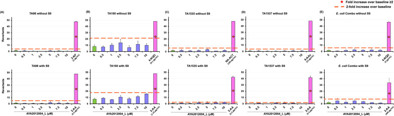
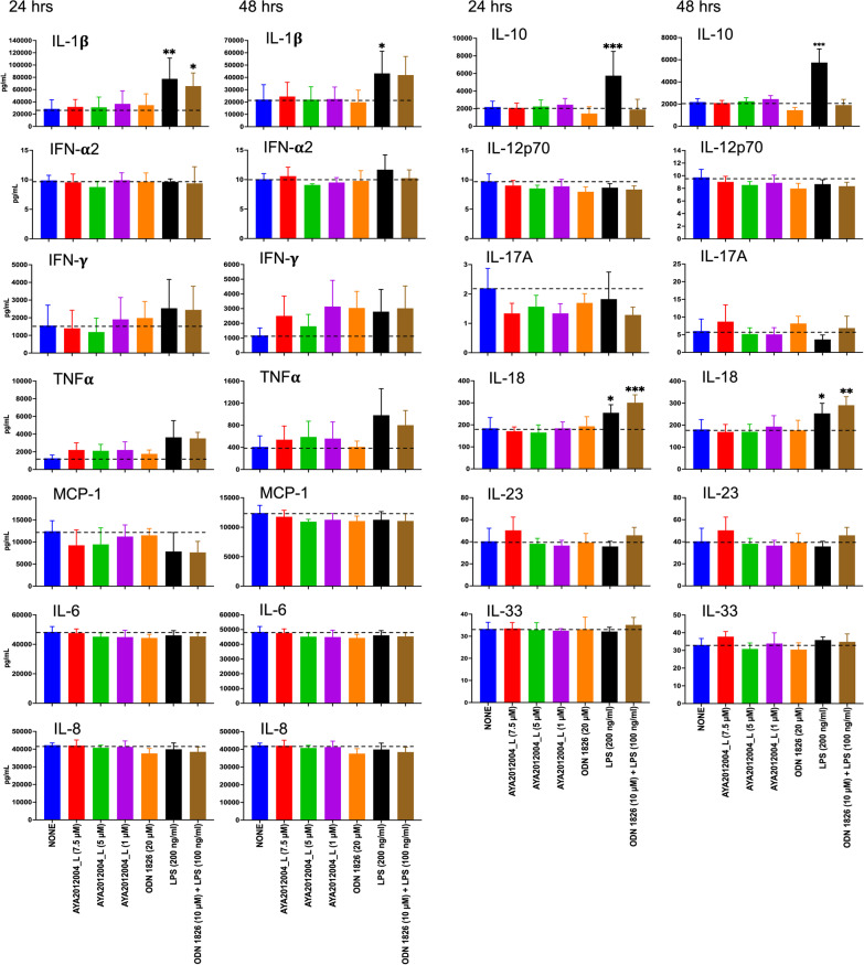
Methods: In vivo studies were conducted in transgenic mice expressing human ACE2 (K18hACE2), C57BL/6J, and Balb/cJ. Flow cytometry was used to check S-protein expressing pseudo-virus-like particles (VLP) uptake by the lung cells and test the immuogenicity of AYA2012004_L. Ames test was used to assess mutagenicity of AYA2012004_L. RT-PCR and histopathology were used to determine the biodistribution and toxicity of AYA2012004_L in vital organs of mice.

Results: We measured the in vivo uptake of VLPs by lung cells by detecting GFP signal using flow cytometry. AYA2012004_L specifically neutralized VLP uptake and also showed no inflammatory response in mice lungs. In addition, AYA2012004_L did not induce inflammatory response in the lungs of Th1 and Th2 mouse models as well as human PBMCs. AYA2012004_L was detectable in mice lungs and noticeable in insignificant amounts in other vital organs. Accumulation of AYA2012004_L in organs decreased over time. AYA2012004_L did not induce degenerative signs in tissues as seen by histopathology and did not cause changes in the body weight of mice. Ames test also certified that AYA2012004_L is non-mutagenic and proved it to be safe for in vivo studies.

Conclusions: Our aptamer is safe, effective, and can neutralize the uptake of VLPs by lung cells when administered locally suggesting that it can be used as a potential therapeutic agent for COVID-19 management.

Keywords: ACE2; Aptamer; COVID-19; Neutralization; RBD; SARS-CoV-2; Safety; Spike S protein; Toxicity; VLP; Virus.


Conflict of interest statement

The authors declare that they have no competing interests.

Figures

Fig. 1
Fig. 1
AYA2012004_L specifically neutralizes S protein VLP uptake by hACE2 transgenic mice lung cells. A Schematic experimental design of mice treatment conditions. B A representative FACS plot showing the neutralization effect of AYA2012004_L on S protein VLP uptake by lung cells. Cells were gated based on DPBS treated control group. C Percent uptake of S protein VLP by lung cells. Uptake of VLPs by lung cells was calculated according to the formula: [(frequency of GFP+ cells of test group − frequency of GFP+ cells of control group)/(frequency of GFP+ cells of VLPs treated group − frequency of GFP+ cells of control group)] × 100. Individual dots represent data generated with cells from different mice. Data are expressed as mean ± SD. The p values were determined with unpaired Student t test. **** denotes p<0.0001
Fig. 2
Fig. 2
Aptamer therapy does not induce inflammatory response in hACE2 transgenic mice lungs. A Schematic experimental design of mice treatment conditions for evaluation of mouse inflammatory cytokines by flow cytometry. B Assessment of cytokines/chemokines from lung cells cultured in the absence or presence of phorbol 12-myristate 13-acetate /ionomycin was done using the LEGENDplex Mouse Inflammation Panel kit and quantitated using flow cytometry. Data were analyzed with BioLegend’s LEGENDplex™ software. Individual dots represent data generated with cells from different mice. The p values were determined with one-way ANOVA, Dunnett’s multiple comparisons test. ns: denotes as nonsignificant
Fig. 3
Fig. 3
Microbial mutagenicity test for AYA2012004_L aptamer’s mutagenic potential is assessed by exposing S. typhimurium strains TA98, TA100, TA1535, TA1537 and E. coli strains wp2[pkM101] and wp2 uvrA to varying concentrations of the aptamer as well as positive and negative controls and selecting for the reversion events. AYA2012004_L was used with increasing concentrations from 0.5 to 10 uM. The assay was performed both in presence and absence of metabolic activation, provided by a liver homogenate S9. Data are a representative over three independent experiments. Data are expressed as mean ±SD
Fig. 4
Fig. 4
Concentration of AYA2012004_L in mouse lungs. C57BL/6J mice were treated with 200 ul of DPBS buffer containing 1 mM MgCl2 in the absence or presence of AYA2012004_L (10 uM) via bronchoalveolar lavage. Mice were then sacrificed at 1, 4, 8, 12, 24 and 48 hrs post-treatment; and lungs were collected and processed for nucleic acid isolation. AYA2012004_L was detected in mice lungs by real-time PCR (RT-PCR) assays. Number of aptamer molecules per mg total DNA was calculated as indicated in the inset picture by plotting a standard curve between ΔCt values and total DNA concentration (pM). Data are representative over two independent experiments
Fig. 5
Fig. 5
Histopathological evaluation of tissues from AYA2012004_L treated mice confirmed no toxicity to vital organs. Photomicrograph of lung, brain, kidney, heart, lymph node, spleen and liver from C57BL/6J mice administered with 200 ul of DPBS in the absence or presence of AYA2012004_L or ODN 1826 via bronchoalveolar lavage. Mice were euthanized at day 45 and tissues were harvested and processed for Hematoxylin and Eosin (H &E) staining. Histopathological evaluation of tissues were described in Table 1. Data are representative over three independent experiments
Fig. 6
Fig. 6
AYA2012004_L does not affect body weight of mice. C57BL/6J mice administered with 200 ul of DPBS in the absence or presence of AYA2012004_L and ODN 1826 via bronchoalveolar lavage. Mice body weight were measured one day before treatment (on day 0) and on day 44 (24 hrs before study termination). The data is expressed as a percent value of the normalization to DPBS treated mice. The p values were determined with one-way ANOVA, Tukey’s multiple comparisons test. ns: denotes nonsignificant.
Fig. 7
Fig. 7
Aptamer AYA2012004_L elicits a partial immunogenic response in mice lung cells that disappeared in 48 hrs. Mice lung cells were stimulated in the presence or absence of AYA2012004_L, LPS, ODN 1826 and LPS+ODN 1826 at 37 C for 24 and 48 hr. Cell supernatants were collected and cytokine levels were assessed using the LEGENDplex Mouse Inflammation Panel kit. Soluble analytes were quantified using flow cytometry and analyzed with BioLegend’s LEGENDplex™ software. Data are representative over three independent experiments
Fig. 8
Fig. 8
Aptamer AYA2012004_L does not elicit an immunogenic response in human peripheral blood mononuclear cells (hPBMCs). Human PBMCs were stimulated with or without AYA2012004_L, LPS, ODN 1826 and LPS+ODN 1826 at 37 C for 24 and 48 h. The amount of cytokines secreted from PBMCs were assessed by the LEGENDplex Human Inflammation kit. Soluble analytes were quantified using flow cytometry and analyzed with BioLegend’s LEGENDplex™ software. The p values were determined with one-way ANOVA, Dunnett’s multiple comparison test. * denotes p<0.05, ** denotes p<0.01 and *** denotes p<0.001

References

    1. Martellucci CA, Flacco ME, Cappadona R, Bravi F, Mantovani L, Manzoli L. Sars-cov-2 pandemic: an overview. Adv Biol Regul. 2020;77:100736. doi: 10.1016/j.jbior.2020.100736. - DOI - PMC - PubMed
    1. Ledford H. Hundreds of covid trials could provide a deluge of new drugs. Nature 2022;25–7. - PubMed
    1. Robinson PC, Liew DF, Tanner HL, Grainger JR, Dwek RA, Reisler RB, Steinman L, Feldmann M, Ho L-P, Hussell T, et al. Covid-19 therapeutics: challenges and directions for the future. Proc Natl Acad Sci. 2022;119(15):2119893119. doi: 10.1073/pnas.2119893119. - DOI - PMC - PubMed
    1. Information on covid-19 treatment, prevention and research. U.S. Department of Health and Human Services. https://www.covid19treatmentguidelines.nih.gov/
    1. Islam S, Islam T, Islam MR. New coronavirus variants are creating more challenges to global healthcare system: a brief report on the current knowledge. Clin Pathol. 2022;15:2632010. doi: 10.1177/2632010X221075584. - DOI - PMC - PubMed

Publication types

Substances

Supplementary concepts