

Splenic lymphangiomas as a common indication for splenectomy: a case series with literature review
- PMID: 36582009
- PMCID: PMC9801652
- DOI: 10.1186/s12893-022-01898-0
Splenic lymphangiomas as a common indication for splenectomy: a case series with literature review
Abstract
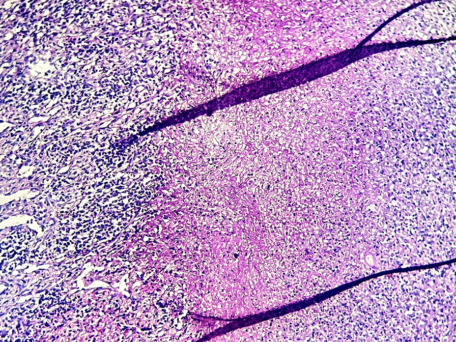
Background: Splenic lymphangiomas (SL) are very rare benign cystic lesions found in pediatric population. Their occurrence in adults is exceptional. Splenectomy is the common management of splenic lesions for diagnostic and/or therapeutic purpose. Our aim is to report additional cases of SL diagnosed on splenectomy specimens at our Pathology laboratory with literature review.
Methods: This is a retrospective study including all cases of splenectomy recorded at our Pathology laboratory (June 2020-August 2022). We performed a comparison of clinicopathological features between patients with SL and those with other benign splenic diseases.
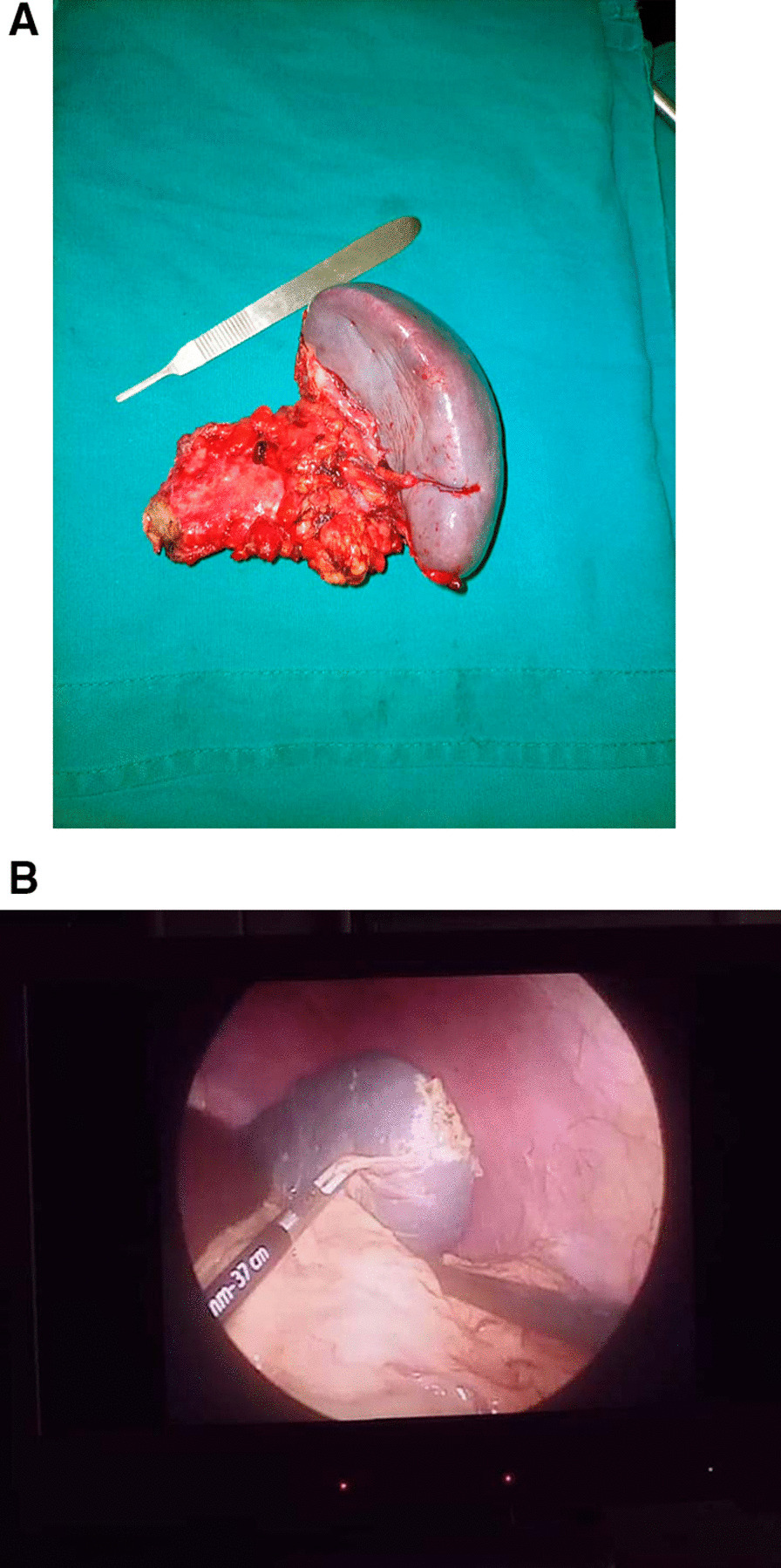
Results: Sixteen cases of splenectomy were included. The mean age was 30.25 years (range of 6-70 years). The final histopathological diagnoses were congestive spleens in all cases of sickle cell disease (SCD) (5/16 patients, 31.25%), splenic cystic lymphangiomas (4/16 patients, 25%), capsular splenic infiltration by gastric and colic cancers (3/16 cases, 18.75%), splenic abscess (2/16 cases, 12.5%) and splenic rupture with subcapsular hematoma (1/16 patients, 6.25%). 12/16 patients (75%) had benign splenic conditions (4/12 with SL, 5/12 with SCD, 2/12 with abscess and 1/12 with splenic trauma). Patients with SL were older than those with other benign splenic conditions (mean age of 28.27 years versus 20.87 years). Also patients with SL presented with massive splenomegaly (mean splenic weight of 1675 g versus 418.75 g, mean splenic size of 19.62 cm versus 14.63 cm). Open surgery was performed in 15/16 patients (93.75%).
Conclusion: Unlike previous studies, our series shows that SL are a common indication for splenectomy and occur in older patients with massive cystic splenomegaly. Open splenectomy is still an usual surgical practice in our country.
Keywords: Cysts; Histopathology; Lymphangioma; Spleen; Splenectomy.
© 2022. The Author(s).
Conflict of interest statement
The authors declare that they have no competing interests.
Figures


Similar articles
-
Redefining the role of splenectomy in patients with idiopathic splenomegaly.ANZ J Surg. 2006 Aug;76(8):679-82. doi: 10.1111/j.1445-2197.2006.03828.x. ANZ J Surg. 2006. PMID: 16916382
-
[Cystic splenic disease of surgical interest].G Chir. 1997 Oct;18(10):555-9. G Chir. 1997. PMID: 9479963 Italian.
-
[Partial splenectomy in non-traumatic benign lesions of the spleen].J Chir (Paris). 1987 May;124(5):326-30. J Chir (Paris). 1987. PMID: 3611233 French.
-
The spectrum of splenic complications in patients with sickle cell disease in Africa: a systematic review.Br J Haematol. 2021 Apr;193(1):26-42. doi: 10.1111/bjh.17179. Epub 2020 Nov 7. Br J Haematol. 2021. PMID: 33161568
-
[Splenic cysts and tumors: diagnosis and management].J Chir (Paris). 2005 Jan-Feb;142(1):6-13. doi: 10.1016/s0021-7697(05)80830-0. J Chir (Paris). 2005. PMID: 15883503 Review. French.
References
-
- Xu G-P, Shen H-F, Yang L-R, Ma Q, Gao B-L. Splenic cystic lymphangioma in a young woman: case report and literature review. Acta Gastroenterol Belg. 2011;74:334–6. - PubMed
Publication types
MeSH terms
LinkOut - more resources
Full Text Sources
Medical
