Review
. 2022 Dec 13:13:1079011.
doi: 10.3389/fimmu.2022.1079011. eCollection 2022.

Steroid receptor coactivators - their role in immunity

Affiliations
Review

Steroid receptor coactivators - their role in immunity

Yosi Gilad et al. Front Immunol. .

Abstract

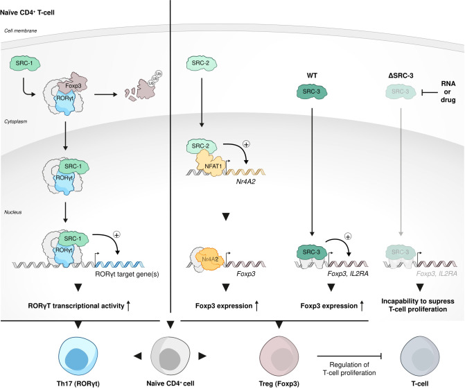
Steroid Receptor Coactivators (SRCs) are essential regulators of transcription with a wide range of impact on human physiology and pathology. In immunology, SRCs play multiple roles; they are involved in the regulation of nuclear factor-κB (NF-κB), macrophage (MΦ) activity, lymphoid cells proliferation, development and function, to name just a few. The three SRC family members, SRC-1, SRC-2 and SRC-3, can exert their immunological function either in an independent manner or act in synergy with each other. In certain biological contexts, one SRC family member can compensate for lack of activity of another member, while in other cases one SRC can exert a biological function that competes against the function of another family counterpart. In this review we illustrate the diverse biological functionality of the SRCs with regard to their role in immunity. In the light of recent development of SRC small molecule inhibitors and stimulators, we discuss their potential relevance as modulators of the immunological activity of the SRCs for therapeutic purposes.

Keywords: Th17 cells; Treg cells; inflammation; macrophages; nuclear coactivators (NCoAs); nuclear factor-κB (NF-κB); steroid receptor coactivators (SRCs).


Conflict of interest statement

The authors are paid consultants by and disclose an equity position in CoRegen, Inc.

Figures

Figure 1
Figure 1
SRCs interactions with NF-κb. (A) SRCs are NF-κB coactivators. SRC-1 can coactivate NF-κB transcriptional activity in an independent manner (top). SRC-1 and SRC-3 cooperate with other coactivators (CBP and p300) for synergistic coactivation of NF-κB (bottom). (B) SRCs drive transcriptional activity of NF-κB under inflammatory-like conditions. SRC-1 interacts with p65 to induce IL-6 transcription under basal and angiotensin II (Ang II)-induced conditions (top). Under TNFα activation, each member of the SRC family (SRC-1, SRC-2 and SRC-3) can drive NF-κB transcriptional activity (bottom).
Figure 2
Figure 2
SRCs role in MΦs. (A) Anti-inflammatory and pro-inflammatory roles of SRC-2 in MΦs. SRC-2 participates in GC-mediated repression of pro-inflammatory cytokine genes: GR-mediated repression of IL1α, IL1β, TNFα, and CCL4 in BMMΦs, following LPS stimulation, positively correlates with co-recruitment of GR and SRC-2 to NF-κB-binding sites occupied by p65 (top). SRC-2 is recruited along with KLF4 to KLF4 target genes in WAT MΦs to promote the induction of anti-inflammatory commitment of these cells (middle left). SRC-2 KO results in attenuated KLF4 expression and activity and drives the shift of WAT-resident MΦs into an inflammatory-like phenotype (middle right). In MG SRC-2 drives neuroinflammation through activation of a proinflammatory program which reflects in a homeostatic signature of MG cells and reduced EAE severity in mice with conditional SRC-2 KO in myeloid cells (bottom). (B) SRC-3 has an anti-inflammatory role in MΦs. SRC-3 stimulation brings about enrichment of anti-inflammatory MΦs: Anti-inflammatory MΦs are enriched by stimulation of SRC-3 with a small molecule stimulator MCB-613 to promote the establishment and maintenance of a pro-reparative environment post MI (top). In colon adenocarcinoma cells SRC-3 interacts with c-Fos to promote KLF4-related gene expression to regulate inflammation (middle). SRC-3 downregulates proinflammatory cytokines: following LPS-stimulation SRC-3 downregulates protein levels, but not mRNA amounts, of proinflammatory cytokines such as TNFα and IL-1β in MΦs, which indicates translational derepression rather than GR-related transcriptional suppression (bottom).
Figure 3
Figure 3
SRCs in Th17 cell development and function. (A) SRC-1-RORγt complex stimulates phenotypic dominance of the Th17 cell lineage over the Treg cell lineage. Following phosphorylation by protein Kinase C θ (PKCθ), SRC-1 replaces FOXP3 in its complex with RORγt in CD4+ cells, subjecting FOXP3 to proteasomal degradation and stimulating Th17 cell lineage phenotypic dominance. (B) SRC-3 is an essential regulator of Th17 cell genes. SRC-3 regulates pathogenic inflammation via coactivation of RORγt-associated expression of Th17 cell genes through an IL-1-ILR1 mediated signaling axis (top). Interaction of RORγt with SRC-3 is important for healthy Th17 cell lineage development: a K313R mutation of RORγt specifically disrupts the ability of RORγt to interact with SRC-3 but not SRC-1 and impairs healthy differentiation and development of Th17 cells (bottom).
Figure 4
Figure 4
SRC-3 role in lymphocyte development. (A) SRC-3 plays an anti-proliferative role in early developing immune cells. SRC-3 selectively inhibits the proliferation of lymphoid cells in a cell autonomous manner: T and B cells isolated from SRC-3 KO mice showed an increased ex vivo proliferation compared to their WT counterparts (left). SRC-3 is required for maintaining the quiescent state of HSCs: lack of SRC-3 results in increased proliferation of HSCs. SRC-3 deficiency in murine HSCs results in an upregulated activity of PGC-1α (the master regulator of mitochondrial biogenesis and metabolism) an consequent disruption of normal HSC function and healthy hematopoiesis (right). (B) SRC-3 is important for healthy development of HSCs, NK and Treg cells. SRC-3 is a critical factor in lineage development and effector function of NK cells: SRC-3 is recruited to the promoter regions of prominent T-bet binding sites in WT but not SRC-3 KO NK cells.
Figure 5
Figure 5
SRC-3 role in Treg cell biology function. Each one of the three SRCs has a distinct role in Treg cell biology and function. SRC-1 replaces FOXP3 in its interaction with RORγt to shift phenotypic dominance of CD4 cells from Treg cell to Th17 cells; SRC-2 stimulates Treg cell differentiation from naïve CD4+ T cells by coactivating NFAT1 to force the expression of a FOXP3 regulator Nr4a2; SRC-3 is enriched and critically important to the biological function of Tregs: inhibition of SRC-3 results in a decrease of transcript levels of the signature Treg cells genes - FOXP3 and IL2RA and consequent incapability of these Tregs to suppress proliferation of conventional T cells.

References

    1. Perissi V, Rosenfeld MG. Controlling nuclear receptors: the circular logic of cofactor cycles. Nat Rev Mol Cell Biol (2005) 6:542–54. doi: 10.1038/nrm1680 - DOI - PubMed
    1. De Bosscher K, Desmet SJ, Clarisse D, Estébanez-Perpiña E, Brunsveld L. Nuclear receptor crosstalk — defining the mechanisms for therapeutic innovation. Nat Rev Endocrinol (2020) 16:363–77. doi: 10.1038/s41574-020-0349-5 - DOI - PubMed
    1. Weikum ER, Liu X, Ortlund EA. The nuclear receptor superfamily: A structural perspective. Protein Sci (2018) 27:1876–92. doi: 10.1002/pro.3496 - DOI - PMC - PubMed
    1. Lonard DM, O'Malley B W. Nuclear receptor coregulators: judges, juries, and executioners of cellular regulation. Mol Cell (2007) 27:691–700. doi: 10.1016/j.molcel.2007.08.012 - DOI - PubMed
    1. Rosenfeld MG, Glass CK. Coregulator codes of transcriptional regulation by nuclear receptors. J Biol Chem (2001) 276:36865–8. doi: 10.1074/jbc.R100041200 - DOI - PubMed

Publication types