

SOX2-positive retinal stem cells are identified in adult human pars plicata by single-cell transcriptomic analyses
- PMID: 36582303
- PMCID: PMC9790047
- DOI: 10.1002/mco2.198
SOX2-positive retinal stem cells are identified in adult human pars plicata by single-cell transcriptomic analyses
Abstract
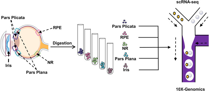
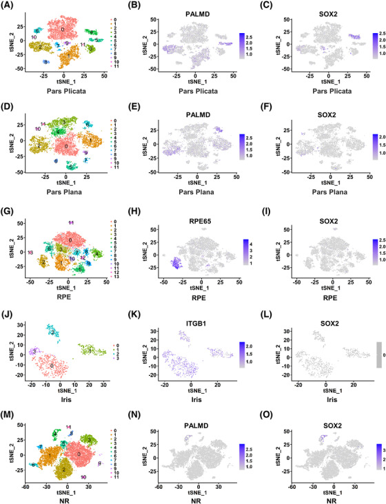
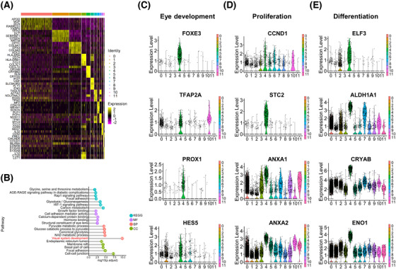
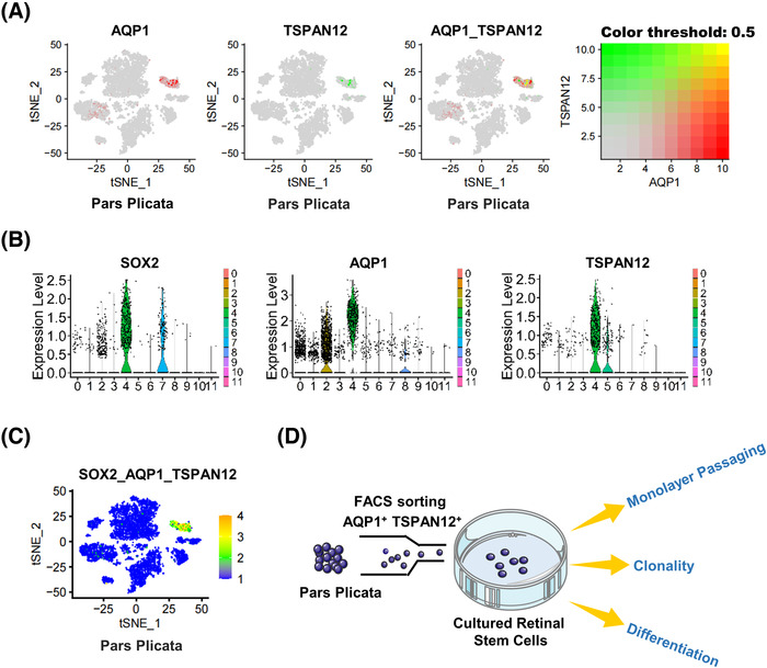
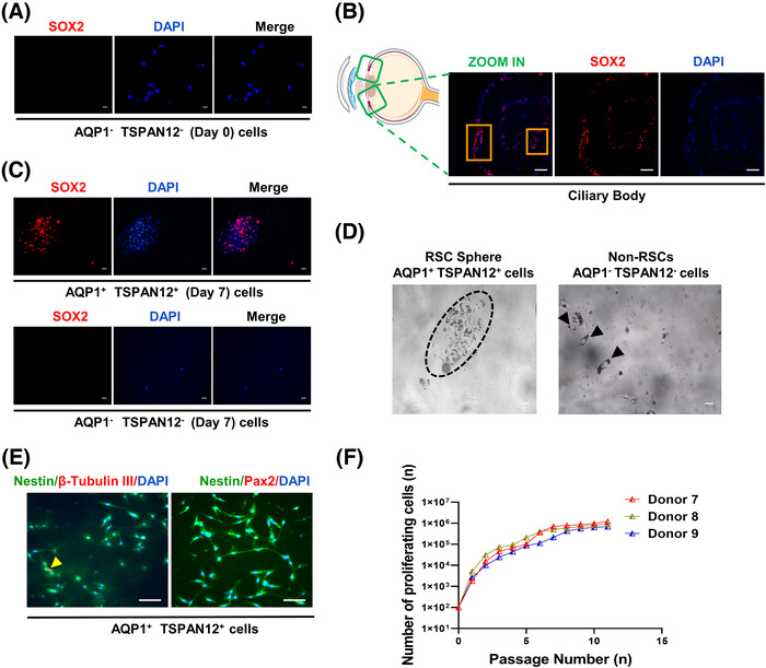
Stem cell therapy is a promising strategy to rescue visual impairment caused by retinal degeneration. Previous studies have proposed controversial theories about whether in situ retinal stem cells (RSCs) are present in adult human eye tissue. Single-cell RNA sequencing (scRNA-seq) has emerged as one of the most powerful tools to reveal the heterogeneity of tissue cells. By using scRNA-seq, we explored the cell heterogeneity of different subregions of adult human eyes, including pars plicata, pars plana, retinal pigment epithelium (RPE), iris, and neural retina (NR). We identified one subpopulation expressing SRY-box transcription factor 2 (SOX2) as RSCs, which were present in the pars plicata of the adult human eye. Further analysis showed the identified subpopulation of RSCs expressed specific markers aquaporin 1 (AQP1) and tetraspanin 12 (TSPAN12). We, therefore, isolated this subpopulation using these two markers by flow sorting and found that the isolated RSCs could proliferate and differentiate into some retinal cell types, including photoreceptors, neurons, RPE cells, microglia, astrocytes, horizontal cells, bipolar cells, and ganglion cells; whereas, AQP1- TSPAN12- cells did not have this differentiation potential. In conclusion, our results showed that SOX2-positive RSCs are present in the pars plicata and may be valuable for treating human retinal diseases due to their proliferation and differentiation potential.
Keywords: SOX2+AQP1+TSPAN12+; retinal degeneration; retinal stem cell; scRNA‐seq.
© 2022 The Authors. MedComm published by Sichuan International Medical Exchange & Promotion Association (SCIMEA) and John Wiley & Sons Australia, Ltd.
Conflict of interest statement
The authors declare that they have no conflict of interest.
Figures

Similar articles
-
Facile isolation and the characterization of human retinal stem cells.Proc Natl Acad Sci U S A. 2004 Nov 2;101(44):15772-7. doi: 10.1073/pnas.0401596101. Epub 2004 Oct 25. Proc Natl Acad Sci U S A. 2004. PMID: 15505221 Free PMC article.
-
Isolation of retinal progenitor and stem cells from the porcine eye.Mol Vis. 2007 Jun 29;13:1045-57. Mol Vis. 2007. PMID: 17653049 Free PMC article.
-
Activation of adult mammalian retinal stem cells in vivo via antagonism of BMP and sFRP2.Stem Cell Res Ther. 2021 Oct 30;12(1):560. doi: 10.1186/s13287-021-02630-0. Stem Cell Res Ther. 2021. PMID: 34717744 Free PMC article.
-
[Variation of inflammatory reaction of ciliary body--harmony between clinic and basic science].Nippon Ganka Gakkai Zasshi. 2004 Dec;108(12):717-48; discussion 749. Nippon Ganka Gakkai Zasshi. 2004. PMID: 15656085 Review. Japanese.
-
[Retinal pigment epithelial cell transplantation: perspective].Nippon Ganka Gakkai Zasshi. 1996 Dec;100(12):982-1006. Nippon Ganka Gakkai Zasshi. 1996. PMID: 9022310 Review. Japanese.
Cited by
-
Single-cell RNA sequencing of retina revealed novel transcriptional landscape in high myopia and underlying cell-type-specific mechanisms.MedComm (2020). 2023 Sep 20;4(5):e372. doi: 10.1002/mco2.372. eCollection 2023 Oct. MedComm (2020). 2023. PMID: 37746666 Free PMC article.
References
-
- Botto C, Rucli M, Tekinsoy MD, Pulman J, Sahel JA, Dalkara D. Early and late stage gene therapy interventions for inherited retinal degenerations. Prog Retin Eye Res. 2022;86:100975. - PubMed
-
- Calabrese EJ. Hormesis and adult adipose‐derived stem cells. Pharmacol Res. 2021;172:105803. - PubMed
-
- Huch M, Koo BK. Modeling mouse and human development using organoid cultures. Development. 2015;142(18):3113‐3125. - PubMed
LinkOut - more resources
Full Text Sources
Miscellaneous