Case Reports
. 2022 Dec 16:46:102304.
doi: 10.1016/j.eucr.2022.102304. eCollection 2023 Jan.

Ancient retroperitoneal schwannoma imitating seminoma recurrence: A case report

Affiliations
Case Reports

Ancient retroperitoneal schwannoma imitating seminoma recurrence: A case report

Samuel Horak et al. Urol Case Rep. .

Abstract

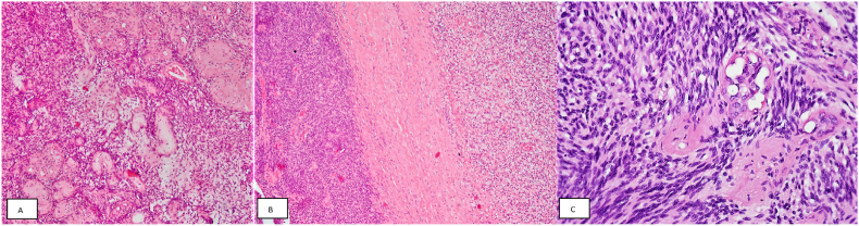
A 28-year-old male with bilateral testicular seminoma underwent bilateral orchiectomy and radiation therapy of the retroperitoneum. After 17 years, he had a retroperitoneal tumor detected, which was removed 7 years later at age 52 because of its progressive enlargement. Due to its partially cystic and partially solid structure, the radiologic findings could not exclude the possibility of regressively altered seminoma metastasis. After radical surgical removal of the tumor, the histopathological and immunohistochemical examination of the tumor revealed ancient schwannoma. These tumors, although unusual, might pose a clinical diagnostic challenge with the risk of undesired overtreatment.

Keywords: Ancient schwannoma; Germ cell tumor; Retroperitoneal; Seminoma metastasis.


Conflict of interest statement

The authors declare no conflict of interests.

Figures

Fig. 1
Fig. 1
PET, CT and PET/CT examination findings (upper row – 17 years after orchiectomy; bottom row – 24 years after orchiectomy). Arrows point towards the enlarging cystic mass in the left retroperitoneum, near the left psoas major muscle.
Fig. 2
Fig. 2
Resected specimen with cystic part (black arrows) and solid part (blue arrows) after grossing. (For interpretation of the references to colour in this figure legend, the reader is referred to the Web version of this article.)
Fig. 3
Fig. 3
A Markedly thickened hyalinized vessels surrounded by tumor cells (HE, 100x). B Hypercellular area (Antoni A – left) and hypocellular area with edematous stroma (Antoni B – right) separated by broad, dense connective tissue band (HE, 100x). C Spindle-shaped tumor cells with bland nuclei, without cytologic or nuclear atypia, growing in bundles in Antoni A area (HE, 400x).

References

    1. Beyer J., Collette L., Sauvé N., et al. International germ cell cancer classification update consortium. Survival and new prognosticators in metastatic seminoma: results from the IGCCCG-update consortium. J Clin Oncol. 2021 May 10;39(14):1553–1562. - PMC - PubMed
    1. Choudry H.A., Nikfarjam M., Liang J.J., et al. Diagnosis and management of retroperitoneal ancient schwannomas. World J Surg Oncol. 2009 Feb 2;7:12. - PMC - PubMed
    1. Rodriguez F.J., Folpe A.L., Giannini C., Perry A. Pathology of peripheral nerve sheath tumors: diagnostic overview and update on selected diagnostic problems. Acta Neuropathol. 2012 Mar;123(3):295–319. - PMC - PubMed
    1. Hamada K., Ueda T., Higuchi I., et al. Peripheral nerve schwannoma: two cases exhibiting increased FDG uptake in early and delayed PET imaging. Skeletal Radiol. 2005 Jan;34(1):52–57. - PubMed
    1. Blok J.M., Kerst J.M., Vegt E., et al. Sentinel node biopsy in clinical stage I testicular cancer enables early detection of occult metastatic disease. BJU Int. 2019 Sep;124(3):424–430. - PMC - PubMed

Publication types

LinkOut - more resources