. 2022 Dec 23;5(12):17640-17651.
doi: 10.1021/acsanm.2c03409. Epub 2022 Dec 2.

Bonding of Neuropeptide Y on Graphene Oxide for Drug Delivery Applications to the Central Nervous System

Affiliations

Bonding of Neuropeptide Y on Graphene Oxide for Drug Delivery Applications to the Central Nervous System

Giada Cellot et al. ACS Appl Nano Mater. .

Erratum in

Abstract

Nanoscale graphene-based materials (GBMs) enable targeting subcellular structures of the nervous system, a feature crucial for the successful engineering of alternative nanocarriers to deliver drugs and to treat neurodisorders. Among GBMs, graphene oxide (GO) nanoflakes, showing good dispersibility in water solution and being rich of functionalizable oxygen groups, are ideal core structures for carrying biological active molecules to the brain, such as the neuropeptide Y (NPY). In addition, when unconjugated, these nanomaterials have been reported to modulate neuronal function per se. Although some GBM-based nanocarriers have been tested both in vitro and in vivo, a thorough characterization of covalent binding impact on the biological properties of the carried molecule and/or of the nanomaterial is still missing. Here, a copper(I)-catalyzed alkyne-azide cycloaddition strategy was employed to synthesize the GO-NPY complex. By investigating through electrophysiology the impact of these conjugates on the activity of hippocampal neurons, we show that the covalent modification of the nanomaterial, while making GO an inert platform for the vectorized delivery, enhances the duration of NPY pharmacological activity. These findings support the future use of GO for the development of smart platforms for nervous system drug delivery.


Conflict of interest statement

The authors declare no competing financial interest.

Figures

Scheme 1
Scheme 1. Functionalization of GO by Epoxide Ring Opening (A), Followed by the Copper Catalyzed Click Reaction (B)
Figure 1
Figure 1
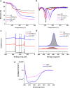
Characterization of the conjugate. (a) TGA of GO, GOTEG-N3, GO–NPY, and NPY, (b) derivative thermogravimetry of GO, GOTEG-N3, GO–NPY, and NPY, (c) XPS survey spectra of GO, GOTEG-N3, and GO–NPY, and (d) XPS high-resolution spectra N 1s of GO, GOTEG-N3, and GO–NPY. (e) CD of NPY and GO–NPY in 1:1 PBS/TFE. In the case of GO–NPY, GOTEG-N3 was used as blanc.
Figure 2
Figure 2
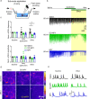
Effect of conjugate on neuronal activity of hippocampal cultures in acute applications. (a) Representative fluorescence microscopy images of hippocampal cultured cells showing the labeling for β-tubulin-positive neurons (in red) and for neuropeptide Y receptors (in green) type 1 (NPY1R; left) and type 2 (NPY2R; right), respectively. DAPI-positive nuclei (in blue). For each image, the area in the white squares was magnified in the upper right insets to better depict the somatic localization of receptors. Scale bar: 20 and 5 μm. (b) Experimental setting. (c) Exemplificative traces for different treatments (saline in black, GO in red, GO–NPY in green and NPY in blue) before (left column) and after (right column) their puff application. (d) Plots showing normalized sPSC frequency and amplitude for the different treatments. Note that only GO induce a post-application increase in sPSC frequency, *P < 0.05.
Figure 3
Figure 3
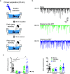
Effect of conjugate on neuronal activity of hippocampal cultures in sub-acute applications. (a) Experimental setting. (b) Exemplificative traces of recordings performed during different treatments (saline in black, GO–NPY in green, and NPY in blue) showing that NPY alone or in conjugation with GO induced a decrease in neuronal activity after some minutes from the beginning of peptide application. (c) Plots showing normalized sPSC frequency and amplitude for the different treatments during the first 3 min of application (early application) and when the effect of the peptide was consolidated (corresponding to the last 2 min of application together with the initial phase of wash out). Note that both GO–NPY and NPY induced a similar decrease in sPSC frequency. (d) GCaMP7f recordings with fluorescence levels in false colors during the baseline and effect periods (top and bottom panels, respectively) for the different conditions. (e) Fluorescence transient traces recorded for the different treatments (saline in black, GO–NPY in green, and NPY in blue) during the baseline and the effects periods. Note that NPY alone or in conjugation with GO induced a decrease in the occurrence of calcium transient after its administration (calcium transient frequencies were 0.017 and 0.019 Hz in control, 0.028 and 0.014 Hz in GO–NPY, and 0.022 and 0.011 Hz in NPY, baseline and effect, respectively). *P < 0.05.
Figure 4
Figure 4
Effect of conjugate on neuronal activity of hippocampal cultures in cronic applications and wash out. (a) Experimental setting. (b) Exemplificative traces of recordings performed after wash out of different treatments (saline in black, GO–NPY in green and NPY in blue) showing that NPY conjugated with GO presented a residual inhibitory action on neuronal activity respect to free NPY. (c) Plots showing sPSC frequency and amplitude for the different treatments during the incubation and after wash out. Note that both GO–NPY and NPY induced a similar decrease in sPSC frequency during the incubation; however, only in cultures exposed to GO–NPY, a subset of neurons presented sPSC frequency lower than 2 Hz after wash out, *P < 0.05.

References

    1. Geim A. K. Graphene: Status and Prospects. Science 2009, 324, 1530–1534. 10.1126/science.1158877. - DOI - PubMed
    1. Kostarelos K.; Novoselov K. S. Exploring the Interface of Graphene and Biology. Science 2014, 344, 261–263. 10.1126/science.1246736. - DOI - PubMed
    1. Sanchez V. C.; Jachak A.; Hurt R. H.; Kane A. B. Biological interactions of graphene-family nanomaterials: An interdisciplinary review. Chem. Res. Toxicol. 2012, 25, 15–34. 10.1021/tx200339h. - DOI - PMC - PubMed
    1. Xia Y. Nanomaterials at work in biomedical research. Nat. Mater. 2008, 7, 758–760. 10.1038/nmat2277. - DOI - PubMed
    1. Silva G. A. Neuroscience nanotechnology: progress, opportunities and challenges. Nat. Rev. Neurosci. 2006, 7, 65–74. 10.1038/nrn1827. - DOI - PubMed