. 2022 Dec 22;15(1):2160536.
doi: 10.1080/20002297.2022.2160536. eCollection 2023.

Ex vivo evaluation of antibiotic sensitivity in samples from endodontic infections

Affiliations

Ex vivo evaluation of antibiotic sensitivity in samples from endodontic infections

Álvaro Villanueva-Castellote et al. J Oral Microbiol. .

Abstract

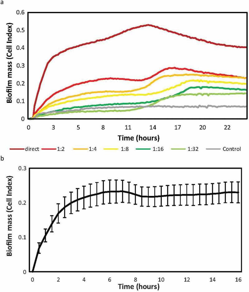
Objective: To develop an in vitro model for real-time monitoring of endodontic biofilm growth and evaluate the ex vivo effect of antibiotics on biofilm growth.

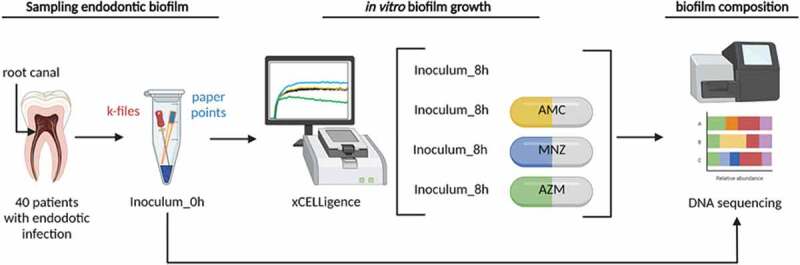
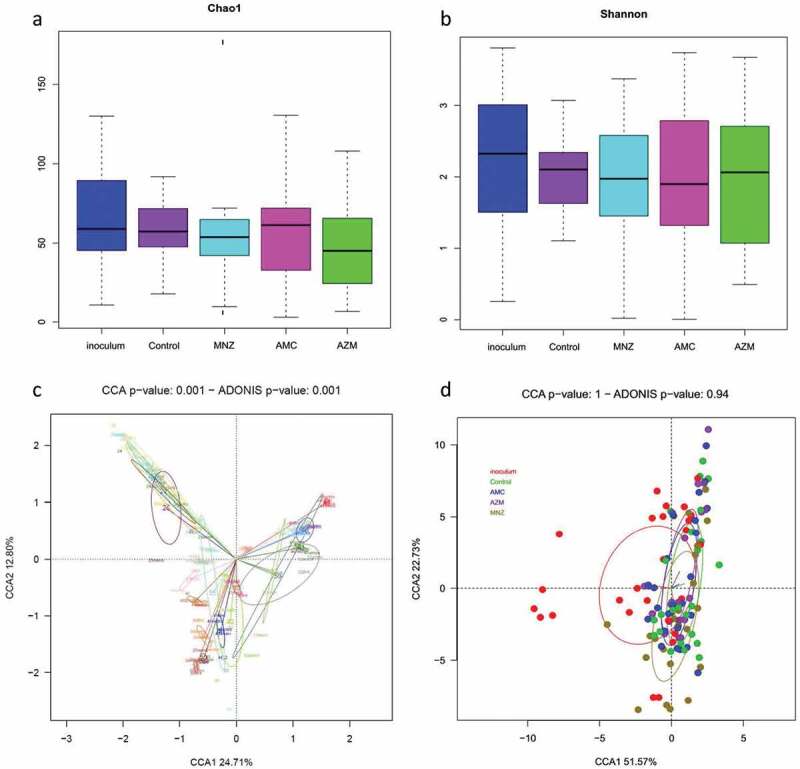
Material and methods: Root canal samples were taken from 40 patients and inoculated into 96-well plates in a system that measures biofilm growth through electrical impedance. Biofilm bacterial composition at the genus and species level was analyzed by Illumina sequencing. ANCOM-BC corrected data were used to compare bacterial composition after antibiotic treatment through compositional analysis, and to compare microbiological with clinical data.

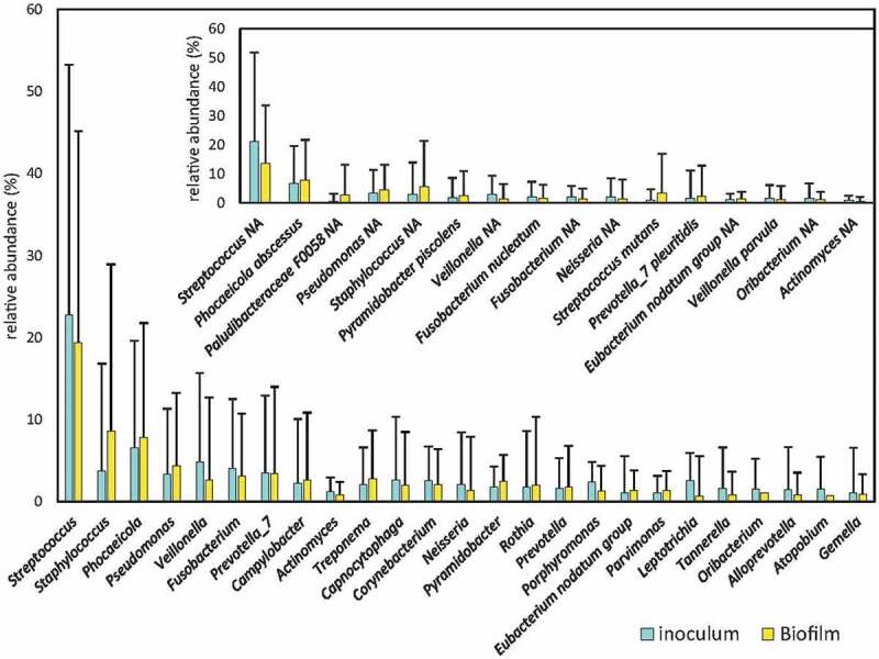
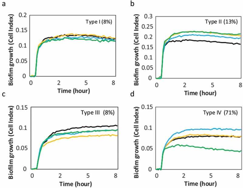
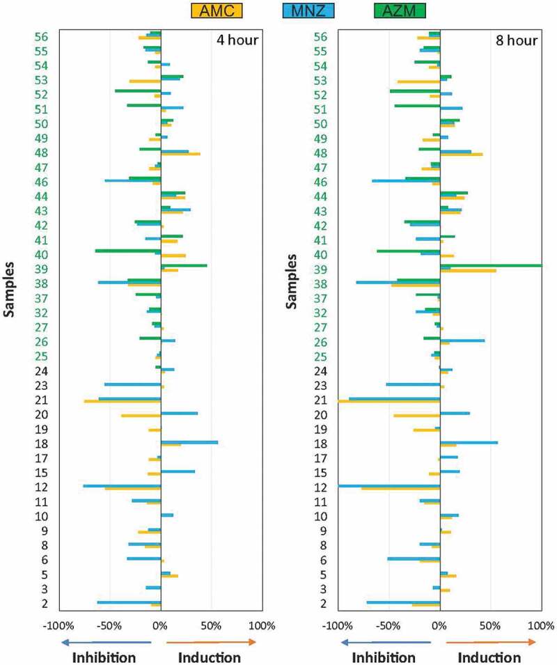
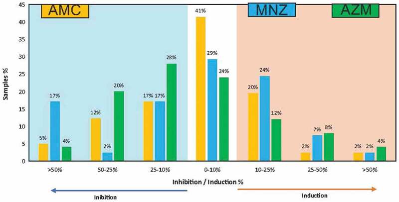
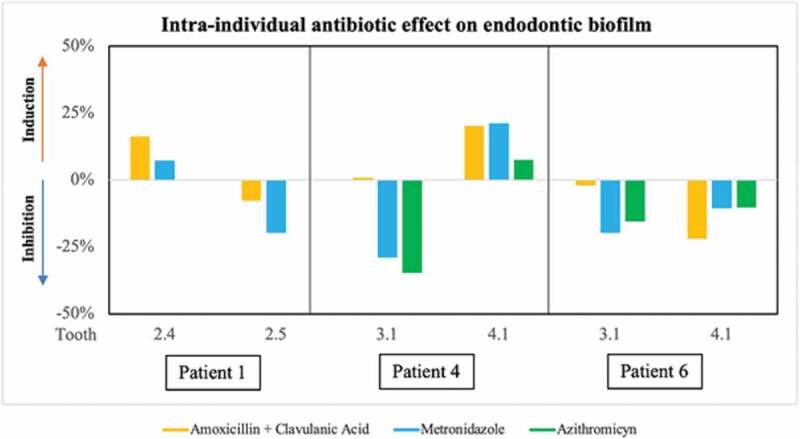
Results: The stationary phase was reached at 8 hours. The biofilm formed had a similar bacterial composition to the inoculum, and Enterococcus faecalis was virtually absent from the samples. The bacterial composition and the effect of antibiotics were sample-dependent. Metronidazole was the antibiotic that most inhibited biofilm formation and azithromycin the one that inhibited it in the highest percentage of cases. The antibiotic effect could not be related to the biofilm original bacterial composition.

Conclusions: The impedance system allowed real-time monitoring of endodontic biofilm formation, and we propose it as a model for ex vivo evaluation of the whole biofilm susceptibility to antimicrobials, as opposed to evaluating antibiotic sensitivity of specific bacterial isolates.

Keywords: Antibiotic; biofilm; biofilm model; endodontic; real time cell analysis.


Conflict of interest statement

No potential conflict of interest was reported by the author(s).

Figures

Figure 1.
Figure 1.
Workflow for microbial analysis of endodontic infections. A schematic view of sample collection, processing, growth and analysis is shown for the antibiotic susceptibility tests, performed with the conditions optimized in the initial set up experiments (1:2 sample dilution and growth for a period of 8 hours). In short, samples from the inside of the necrotic teeth canals were obtained, and the files and paper points with the sample were placed in an Eppendorf tube with PBS. An aliquot was taken from each sample and used for the study of microbial composition by 16S rRNA gene Illumina sequencing. Samples were also inoculated into the wells of E-plates from a xCelligence® RTCA system and incubated at 37°C for 8 hours to study the dynamics of endodontic biofilm formation. Each well contains 100 µl of culture medium with each of the three antibiotics tested (AMC- medium with amoxicillin-clavulanic acid, MNZ- medium with metronidazole and AZM- medium with azithromycin), 100 µl of sample cell suspension (inoculum) and 50 µl of sterile mineral oil to facilitate the creation of anaerobic conditions. Once the biofilm reached the stationary phase (8 hours), the biofilm attached to the bottom of the well was collected for DNA extraction and 16S rRNA gene sequencing, which was carried out using Illumina Miseq followed by bioinformatics analysis with compositional analysis.
Figure 2.
Figure 2.
Endodontic biofilm formation dynamics after serial dilutions. a) the y-axis indicates the impedance values (i.e. biofilm growth) measured over 24 h. Sample was diluted from an initial concentration 1/1 (pellet diluted in 500 ul of medium), up to a dilution 1/32. The results show the mean Cell Index (CI) or biofilm growth values of two replicates recorded in real time over 24 h of biofilm formation on an RTCA xCelligence system. The grey line corresponds to the negative control of the medium without sample. b) Mean and SE (standard error) of endodontic biofilm growth of 9 samples of different patients, measured in real time for 16 h on the RTCA xCelligence® system. For each sample, the mean of the 2 replicates was taken and the impedance values were subtracted from the blank (control medium).
Figure 3.
Figure 3.
Bacterial composition of root canal infections. Graphs show the mean proportion and SE (standard error) of the most abundant genera in the samples (those at >1% levels) from inside the root canal (inoculum) and the derived biofilms grown ex vivo. Inlet graph shows the proportion of the 15 most abundant bacterial species in the samples. * (p < 0.05).
Figure 4.
Figure 4.
Patterns of ex vivo growth of endodontic biofilms derived from necrotic root canals under different antibiotics. The curves show the biofilm growth measured in real time on the xCelligence® RTCA system in the absence of antibiotics (positive control, black curve) or in the presence of antibiotics: amoxicillin + clavulanic acid (yellow), metronidazole (blue), azithromycin (green). Panels a, b, c and d show representative examples of the effect on biofilm formation from each of the 4 patterns found in response to the antibiotics. The percentage frequency of the different biofilm growth patterns found in the 40 samples studied are indicated between brackets. -Type I: no effect; Type II: all antibiotics induce; Type III: all antibiotics inhibit; Type IV: variable effects for different antibiotics-.
Figure 5.
Figure 5.
Graphical representation of the percentage of inhibition or induction of biofilm formation in vitro. Bar graphs show data from 40 samples of endodontic origin, grown in the presence of different antibiotics, at 4 and 8 hours, compared to a control without antibiotics.
Figure 6.
Figure 6.
Inhibition or induction frequency of biofilm formation for the different antibiotics tested ex vivo. Bar graphs show data from 40 endodontic samples grown for 8 hours. The bars express the percentage of samples in which each antibiotic induced or inhibited biofilm formation with respect to the control sample without antibiotics, for different degrees of efficacy.
Figure 7.
Figure 7.
Microbial composition analyses of endodontic infections and their corresponding biofilms grown in the absence and presence of antibiotics. The analysis was performed on all patient samples from which endodontic biofilm grown was obtained in the absence of antibiotics (control) and after the application of amoxicillin + clavulanic acid (AMC), metronidazole (MNZ) and azithromycin (AZM). a) Bacterial richness (estimated number of bacterial species) as determined by the Chao1 index. c) Bacterial diversity as determined by the Shannon index. Lower panels represent a graphical representation by canonical correspondence analysis (CCA) of the microbial community composition of the biofilm as grouped by patient (c) and by antibiotic treatment (d). In panel (c), each circle and color corresponds to a different individual. Each sample occupies a position in the 2-D space according to its bacterial composition at the species level as determined by 16S rRNA sequencing.
Figure 8.
Figure 8.
Graphical representation of the effect of the antibiotics tested ex vivo on biofilm formation from two samples obtained from each of 3 patients. Data are shown from patients 1, 4 and 6, shown as percentage of inhibition or induction of biofilm formation relative to the control sample without antibiotics at 8 h of growth. The bars show the mean of two replicates.

Similar articles

Cited by

References

    1. Alves FR, Almeida BM, Neves MA, et al. Disinfecting oval-shaped root canals: effectiveness of different supplementary approaches. J Endod. 2013;37(4):496–14. DOI: 10.1016/j.joen.2010.12.008 - DOI - PubMed
    1. Siqueira Jr JF, Roças IN.. Optimising single-visit disinfection with supplementary approaches: a quest for predictability. Aust Endod J. 2011;37(3):92–99. - PubMed
    1. Ricucci D, Siqueira JF. Biofilms and apical periodontitis: study of prevalence and association with clinical and histopathologic findings. J Endod. 2010;36(8):1277–1288. - PubMed
    1. Provenzano JC, Siqueira JF, Roças IN, et al. Metaproteome analysis of endodontic infections in association with different clinical conditions. PLoS ONE. 2013;8(10):e76108. DOI: 10.1371/journal.pone.0076108 - DOI - PMC - PubMed
    1. Neelakantan P, Romero M, Vera J, et al. Biofilms in Endodontics—Current status and future directions. Int J Mol Sci. 2017;18(8):1748. DOI: 10.3390/ijms18081748 - DOI - PMC - PubMed

LinkOut - more resources