

Retinal Vascular Geometry in Hypertension: cSLO-Based Method
- PMID: 36583807
- PMCID: PMC10011349
- DOI: 10.1007/s40123-022-00642-4
Retinal Vascular Geometry in Hypertension: cSLO-Based Method
Abstract
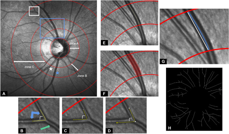
Introduction: We aim to introduce a method using confocal scanning laser ophthalmoscopy (cSLO) images for measuring retinal vascular geometry, including vessel branch angle (BA), vessel diameter, vessel tortuosity, and fractal dimension (Df), and to elucidate the relationship between hypertension and these metrics.
Methods: A total of 119 participants (119 eyes) were enrolled, among which 72 were normotensive and 47 were hypertensive. Infrared cSLO images were extracted from the circular scan around the optics disc using a commercial cSLO + optical coherence tomography instrument. Preprocessed cSLO images were further analyzed using the appropriate tool/macro/plugin of ImageJ.
Results: Intraclass correlation coefficients of selected methods used for conducting the cSLO-based geometric analyses were all higher than 0.80. Arterial/arteriolar BA, arteriolar vessel diameter, and total Df in normotensive subjects were 85.80 ± 7.79°, 116.80 ± 12.58 μm, and 1.430 ± 0.037, respectively, significantly higher than those of hypertensive subjects (82.13 ± 10.83°, 108.2 ± 11.12 μm, and 1.361 ± 0.044, all P < 0.05). The aforementioned metrics remained negatively correlated with hypertension even after adjusting for age alone or age and gender (P < 0.05). However, the difference between arteriolar tortuosity and all studied venous/venular geometric parameters in both subjects was insignificant (all P > 0.05).
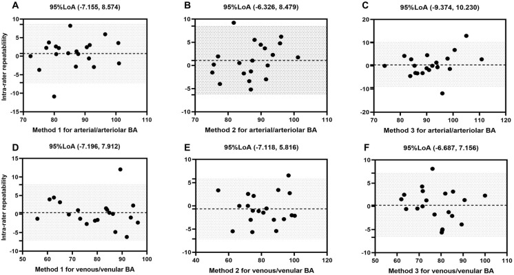
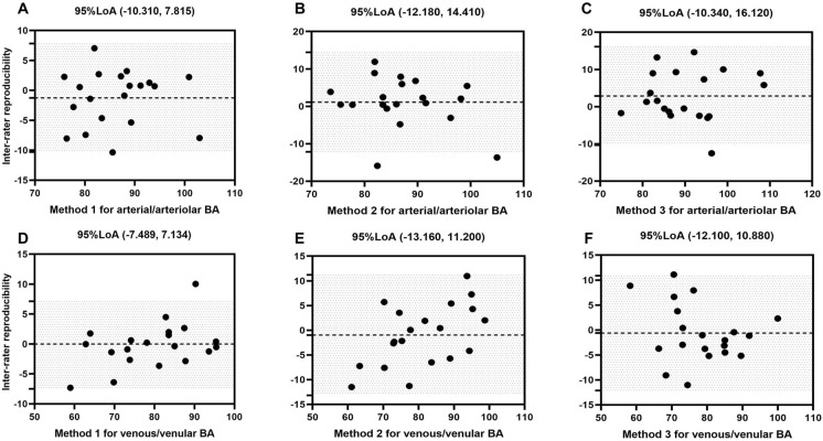
Conclusion: Proposed cSLO-based methods for assessing various vascular geometric parameters were highly repeatable and reproducible. Arterial/arteriolar BA, arteriolar vessel diameter, and total Df were retinal vascular parameters significantly correlated with hypertension in a negative manner.
Keywords: OCT; Retinal vascular geometry; cSLO.
© 2022. The Author(s).
Figures

Similar articles
-
Comparison of confocal scanning laser ophthalmoscopy, scanning laser polarimetry and optical coherence tomography to discriminate ocular hypertension and glaucoma at an early stage.Graefes Arch Clin Exp Ophthalmol. 2006 Jan;244(1):58-68. doi: 10.1007/s00417-005-0029-0. Epub 2005 Jul 26. Graefes Arch Clin Exp Ophthalmol. 2006. PMID: 16044326
-
Comparison of retinal nerve fiber layer imaging by spectral domain optical coherence tomography and scanning laser ophthalmoscopy.Ophthalmology. 2011 Nov;118(11):2196-202. doi: 10.1016/j.ophtha.2011.03.035. Epub 2011 Jul 16. Ophthalmology. 2011. PMID: 21762989
-
Longitudinal evaluation of optic disc measurement variability with optical coherence tomography and confocal scanning laser ophthalmoscopy.J Glaucoma. 2009 Feb;18(2):101-6. doi: 10.1097/IJG.0b013e318179f879. J Glaucoma. 2009. PMID: 19225344
-
Evaluation of the influence of tilt of optic disc on the measurement of optic disc variables obtained by optical coherence tomography and confocal scanning laser ophthalmoscopy.J Glaucoma. 2005 Jun;14(3):210-4. doi: 10.1097/01.ijg.0000159129.93085.96. J Glaucoma. 2005. PMID: 15870603
-
Retinal Vascular Signs and Cerebrovascular Diseases.J Neuroophthalmol. 2020 Mar;40(1):44-59. doi: 10.1097/WNO.0000000000000888. J Neuroophthalmol. 2020. PMID: 31977663 Review.
References
-
- Koch E, Rosenbaum D, Brolly A, Sahel JA, Chaumet-Riffaud P, Girerd X, et al. Morphometric analysis of small arteries in the human retina using adaptive optics imaging: relationship with blood pressure and focal vascular changes. J Hypertens. 2014;32:890–898. doi: 10.1097/HJH.0000000000000095. - DOI - PMC - PubMed
-
- Forster RB, Garcia ES, Sluiman AJ, Grecian SM, McLachlan S, MacGillivray TJ, et al. Retinal venular tortuosity and fractal dimension predict incident retinopathy in adults with type 2 diabetes: the Edinburgh type 2 diabetes study. Diabetologia. 2021;64:1103–1112. doi: 10.1007/s00125-021-05388-5. - DOI - PMC - PubMed
Grants and funding
LinkOut - more resources
Full Text Sources
Miscellaneous
