Type I Interferon Signaling Is Critical During the Innate Immune Response to HSV-1 Retinal Infection
- PMID: 36583876
- PMCID: PMC9807183
- DOI: 10.1167/iovs.63.13.28
Type I Interferon Signaling Is Critical During the Innate Immune Response to HSV-1 Retinal Infection
Abstract
Purpose: Acute retinal necrosis (ARN) is a herpesvirus infection of the retina with blinding complications. In this study, we sought to create a reproducible mouse model of ARN that mimics human disease to better understand innate immunity within the retina during virus infection.
Methods: C57Bl/6J wild type (WT) and type I interferon receptor-deficient (IFNAR-/-) mice were infected with varying amounts of herpes simplex virus type 1 (HSV-1) via subretinal injection. Viral titers, optical coherence tomography (OCT) and fundus photography, the development of encephalitis, and ocular histopathology were scored and compared between groups of WT and IFNAR-/- mice.
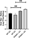
Results: The retina of WT mice could be readily infected with HSV-1 via subretinal injection resulting in retinal whitening and full-thickness necrosis as determined by in vivo imaging and histopathology. In IFNAR-/- mice, HSV-1-induced retinal pathology was significantly worse when compared with WT mice, and viral titers were significantly elevated within two days after infection and persisted to day 5 after infection within the retina. These results were also observed in the brain where there were significantly higher viral titers and frequency of encephalitis in IFNAR-/- when compared to WT mice.
Conclusions: Collectively, these findings show that our new mouse model of ARN mimics human disease and can be used to study innate immunity within the retina. We conclude that type I interferons are critical in containing HSV-1 locally within retinal tissues and prohibiting spread into the brain.
Conflict of interest statement
Disclosure:
Figures


Similar articles
-
The Type I Interferon Response Determines Differences in Choroid Plexus Susceptibility between Newborns and Adults in Herpes Simplex Virus Encephalitis.mBio. 2016 Apr 12;7(2):e00437-16. doi: 10.1128/mBio.00437-16. mBio. 2016. PMID: 27073094 Free PMC article.
-
Immune- and Nonimmune-Compartment-Specific Interferon Responses Are Critical Determinants of Herpes Simplex Virus-Induced Generalized Infections and Acute Liver Failure.J Virol. 2016 Nov 14;90(23):10789-10799. doi: 10.1128/JVI.01473-16. Print 2016 Dec 1. J Virol. 2016. PMID: 27681121 Free PMC article.
-
The Innate Immune Response to Herpes Simplex Virus 1 Infection Is Dampened in the Newborn Brain and Can Be Modulated by Exogenous Interferon Beta To Improve Survival.mBio. 2020 May 26;11(3):e00921-20. doi: 10.1128/mBio.00921-20. mBio. 2020. PMID: 32457247 Free PMC article.
-
Acute retinal necrosis caused by herpes simplex virus type 2 in children: reactivation of an undiagnosed latent neonatal herpes infection.Semin Pediatr Neurol. 2012 Sep;19(3):115-8. doi: 10.1016/j.spen.2012.02.005. Semin Pediatr Neurol. 2012. PMID: 22889540 Free PMC article. Review.
-
Evasion of host antiviral innate immunity by HSV-1, an update.Virol J. 2016 Mar 8;13:38. doi: 10.1186/s12985-016-0495-5. Virol J. 2016. PMID: 26952111 Free PMC article. Review.
Cited by
-
Immunological Considerations for the Development of an Effective Herpes Vaccine.Microorganisms. 2024 Sep 6;12(9):1846. doi: 10.3390/microorganisms12091846. Microorganisms. 2024. PMID: 39338520 Free PMC article. Review.
-
Case Reports: Chemokine and Cytokine Profiling in Patients with Herpetic Uveitis.Int Med Case Rep J. 2024 Dec 19;17:1055-1061. doi: 10.2147/IMCRJ.S496941. eCollection 2024. Int Med Case Rep J. 2024. PMID: 39719962 Free PMC article.
-
Infectious Eye Diseases and Prevention Control.Microorganisms. 2023 May 15;11(5):1286. doi: 10.3390/microorganisms11051286. Microorganisms. 2023. PMID: 37317260 Free PMC article.
-
A Better Understanding of the Clinical and Pathological Changes in Viral Retinitis: Steps to Improve Visual Outcomes.Microorganisms. 2024 Dec 5;12(12):2513. doi: 10.3390/microorganisms12122513. Microorganisms. 2024. PMID: 39770716 Free PMC article. Review.
-
The Host-Pathogen Interplay: A Tale of Two Stories within the Cornea and Posterior Segment.Microorganisms. 2023 Aug 12;11(8):2074. doi: 10.3390/microorganisms11082074. Microorganisms. 2023. PMID: 37630634 Free PMC article. Review.
References
-
- Kopplin LJ, Thomas AS, Cramer S, et al. .. Long-term surgical outcomes of retinal detachment associated with acute retinal necrosis. Opht Surg Lasers Img Retina. 2016; 47: 660–664. - PubMed
-
- Silva RA, Berrocal AM, Moshfeghi DM, Blumenkranz MS, Sanislo S, Davis JL. Herpes simplex virus type 2 mediated acute retinal necrosis in a pediatric population: case series and review. Graefes Arch Clin Exp Ophthalmol. 2013; 251: 559–566. - PubMed
-
- Schoenberger SD, Kim SJ, Thorne JE, et al. .. Diagnosis and treatment of acute retinal necrosis: a report by the American Academy of Ophthalmology. Ophthalmology. 2017; 124: 382–392. - PubMed
-
- Hadden PW, Barry CJ. Herpetic encephalitis and acute retinal necrosis. N Engl J Med. 2002; 347: 1932–1932. - PubMed
Publication types
MeSH terms
Substances
Grants and funding
LinkOut - more resources
Full Text Sources
Medical
Molecular Biology Databases

