

Bi-Directional Axial Transmission measurements applied in a clinical environment
- PMID: 36584002
- PMCID: PMC9803229
- DOI: 10.1371/journal.pone.0277831
Bi-Directional Axial Transmission measurements applied in a clinical environment
Abstract
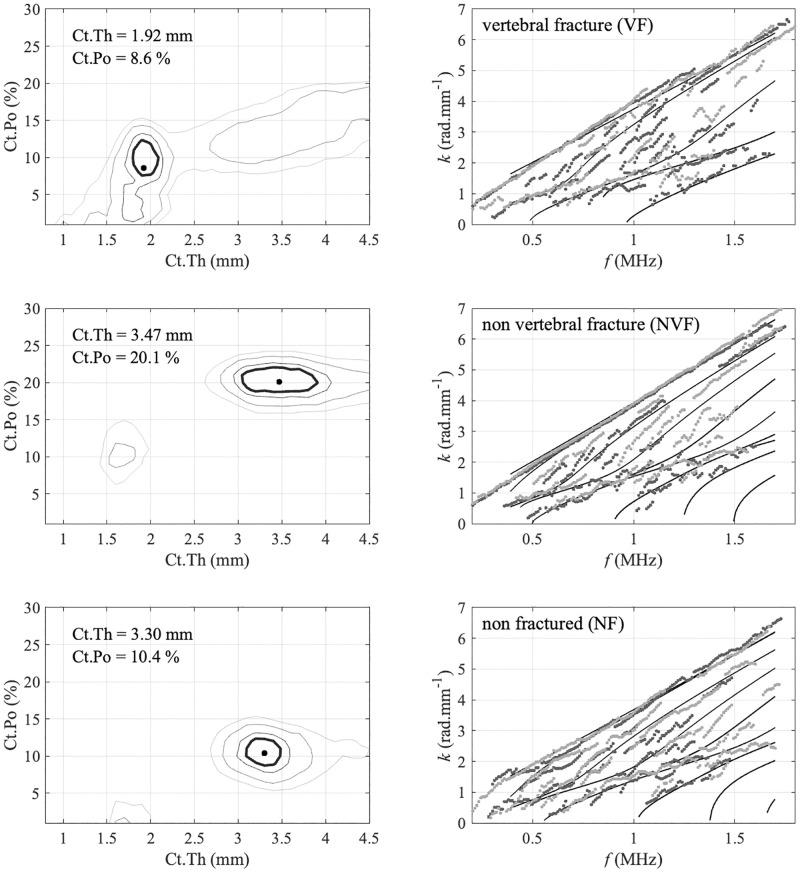
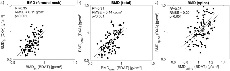
Accurate measurement of cortical bone parameters may improve fracture risk assessment and help clinicians on the best treatment strategy. Patients at risk of fracture are currently detected using the current X-Ray gold standard DXA (Dual XRay Absorptiometry). Different alternatives, such as 3D X-Rays, Magnetic Resonance Imaging or Quantitative Ultrasound (QUS) devices, have been proposed, the latter having advantages of being portable and sensitive to mechanical and geometrical properties. The objective of this cross-sectional study was to evaluate the performance of a Bi-Directional Axial Transmission (BDAT) device used by trained operators in a clinical environment with older subjects. The device, positioned at one-third distal radius, provides two velocities: VFAS (first arriving signal) and VA0 (first anti-symmetrical guided mode). Moreover, two parameters are obtained from an inverse approach: Ct.Th (cortical thickness) and Ct.Po (cortical porosity), along with their ratio Ct.Po/Ct.Th. The areal bone mineral density (aBMD) was obtained using DXA at the femur and spine. One hundred and six patients (81 women, 25 men) from Marien Hospital and St. Anna Hospital (Herne, Germany) were included in this study. Age ranged from 41 to 95 years, while body mass index (BMI) ranged from 16 to 47 kg.m-2. Three groups were considered: 79 non-fractured patients (NF, 75±13years), 27 with non-traumatic fractures (F, 80±9years) including 14 patients with non-vertebral fractures (NVF, 84±7years). Weak to moderate significant Spearman correlations (R ranging from 0.23 to 0.53, p < 0.05) were found between ultrasound parameters and age, BMI. Using multivariate Partial Least Square discrimination analyses with Leave-One-Out Cross-Validation (PLS-LOOCV), we found the combination of VFAS and the ratio Ct.Po/Ct.Th to be predictive for all non traumatic fractures (F) with the odds ratio (OR) equals to 2.5 [1.6-3.4] and the area under the ROC curve (AUC) equal to 0.63 [0.62-0.65]. For the group NVF, combination of four parameters VA0. Ct.Th, Ct.Po and Ct.Po/Ct.Po, along with age provides a discrimination model with OR and AUC equals to 7.5 [6.0-9.1] and 0.75 [0.73-0.76]. When restricted to a smaller population (87 patients) common to both BDAT and DXA, BDAT ORs and AUCs are comparable or slightly higher to values obtained with DXA. The fracture risk assessment by BDAT method in older patients, in a clinical setting, suggests the benefit of the affordable and transportable device for the routine use.
Copyright: © 2022 Minonzio et al. This is an open access article distributed under the terms of the Creative Commons Attribution License, which permits unrestricted use, distribution, and reproduction in any medium, provided the original author and source are credited.
Conflict of interest statement
The authors state that they have no conflicts of interest.
Figures

References
Publication types
MeSH terms
LinkOut - more resources
Full Text Sources
Medical
