. 2023 Jan 3;120(1):e2217476120.
doi: 10.1073/pnas.2217476120. Epub 2022 Dec 30.

The chaperone protein p32 stabilizes HIV-1 Tat and strengthens the p-TEFb/RNAPII/TAR complex promoting HIV transcription elongation

Affiliations

The chaperone protein p32 stabilizes HIV-1 Tat and strengthens the p-TEFb/RNAPII/TAR complex promoting HIV transcription elongation

Chuan Li et al. Proc Natl Acad Sci U S A. .

Abstract

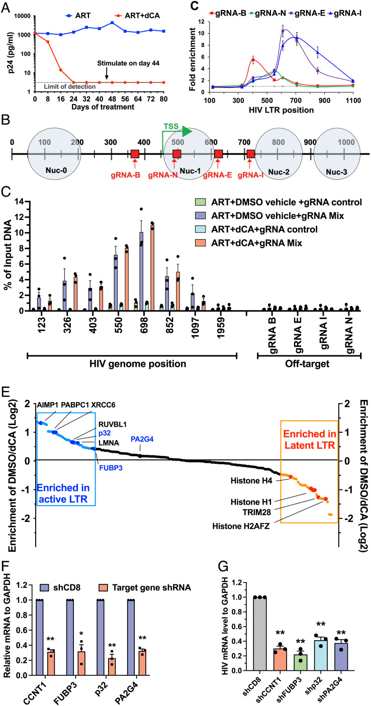
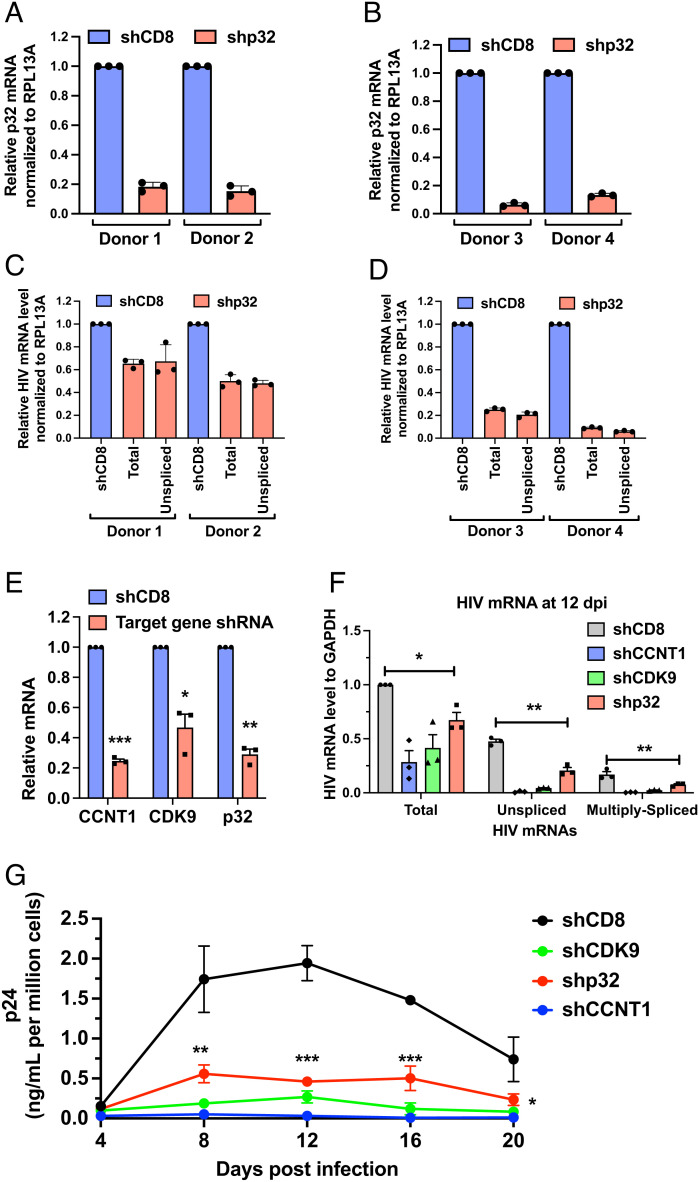
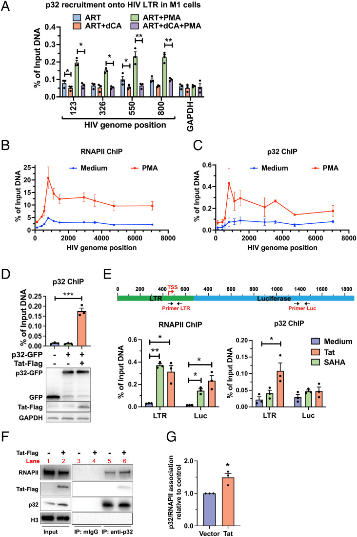
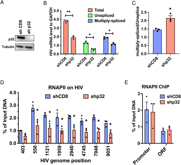
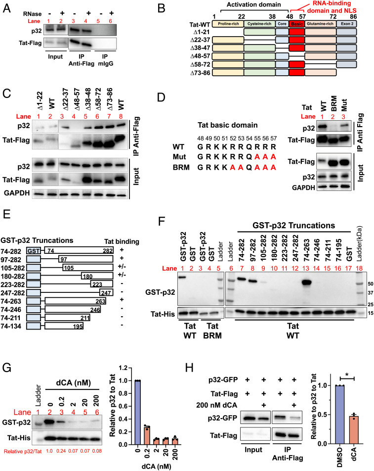
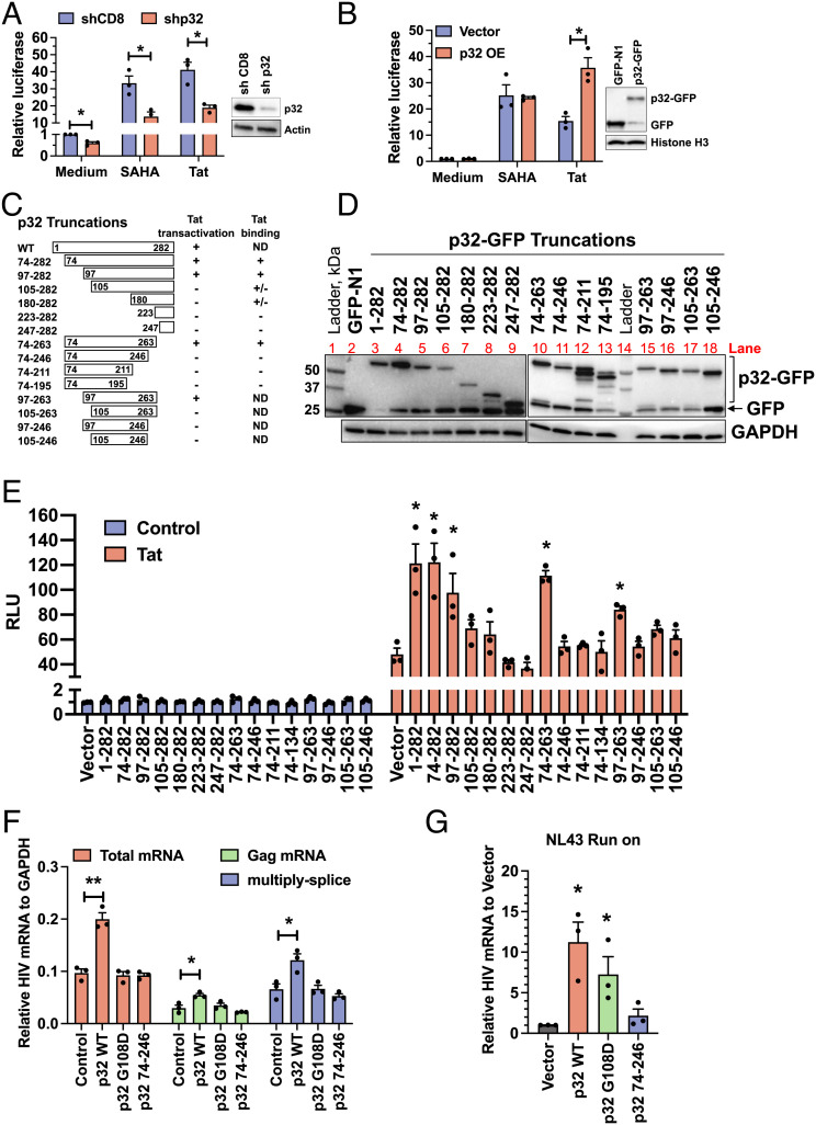
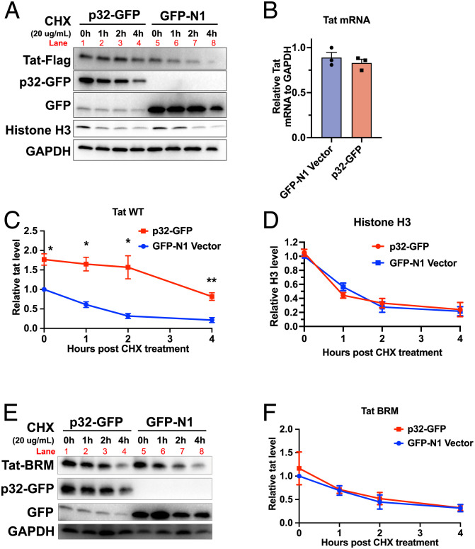
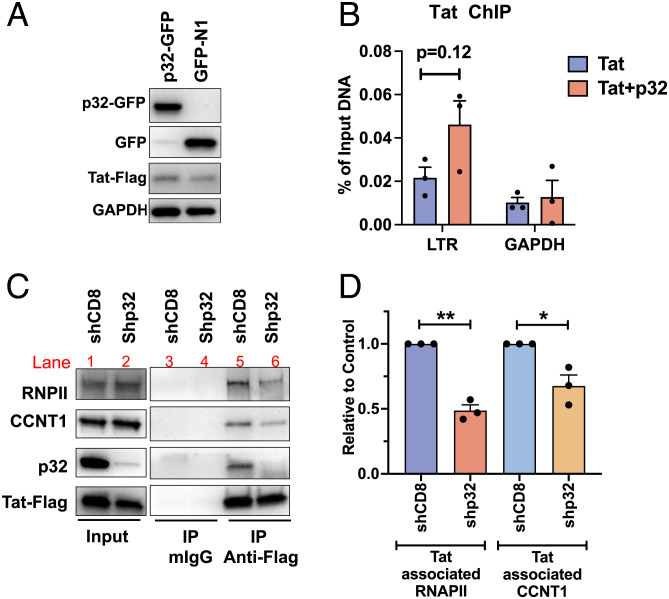
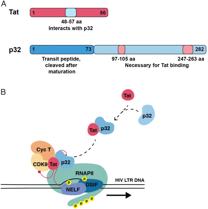
HIV gene expression is modulated by the combinatorial activity of the HIV transcriptional activator, Tat, host transcription factors, and chromatin remodeling complexes. To identify host factors regulating HIV transcription, we used specific single-guide RNAs and endonuclease-deficient Cas9 to perform chromatin affinity purification of the integrated HIV promoter followed by mass spectrometry. The scaffold protein, p32, also called ASF/SF2 splicing factor-associated protein, was identified among the top enriched factors present in actively transcribing HIV promoters but absent in silenced ones. Chromatin immunoprecipitation analysis confirmed the presence of p32 on active HIV promoters and its enhanced recruitment by Tat. HIV uses Tat to efficiently recruit positive transcription elongation factor b (p-TEFb) (CDK9/CCNT1) to TAR, an RNA secondary structure that forms from the first 59 bp of HIV transcripts, to enhance RNAPII transcriptional elongation. The RNA interference of p32 significantly reduced HIV transcription in primary CD4+T cells and in HIV chronically infected cells, independently of either HIV splicing or p32 anti-splicing activity. Conversely, overexpression of p32 specifically increased Tat-dependent HIV transcription. p32 was found to directly interact with Tat's basic domain enhancing Tat stability and half-life. Conversely, p32 associates with Tat via N- and C-terminal domains. Likely due its scaffold properties, p32 also promoted Tat association with TAR, p-TEFb, and RNAPII enhancing Tat-dependent HIV transcription. In sum, we identified p32 as a host factor that interacts with and stabilizes Tat protein, promotes Tat-dependent transcriptional regulation, and may be explored for HIV-targeted transcriptional inhibition.

Keywords: HIV; Tat; p32; regulation; transcription.


Conflict of interest statement

The authors declare no competing interest.

Figures

Fig. 1.
Fig. 1.
Identification of HIV promoter (5′LTR)-associated proteins by ChAP–MS. A. HeLa–M1 cells chronically infected with HIV NL4-3 were treated with ART or ART + dCA (100 nM) over time and viral capsid (p24) production in the supernatant monitored by p24 ELISA. Cells were split on average every 4 d. Data are n=3. B. Schematic of the HIV promoter (5′LTR) region 1 to 1,100 nt. and location of the four gRNAs. Nuc locations are represented by gray circles. TSS (green arrow): Transcription start site. C. HIV promoter enrichment after purification with the indicated gRNAs. Cells were transfected with dCAS9 and a single gRNA and collected 48 h later, crosslinked, and subjected to chromatin purification. HIV-specific primers used to quantify HIV promoter enrichment by qPCR. Fold enrichment: % of input DNA of a specific gRNA divided by % of input DNA of gRNA to GFP (negative control). Data represent mean ± STE (n=3). D. HIV promoter DNA enrichment in both ART + DMSO and ART + dCA-treated M1. Cells were transfected with dCAS9 and a mixture of gRNAs B, N, E, and I or control gRNA to GFP. HIV-specific primers used to analyze HIV promoter enrichment by qPCR. Enrichment of similar sites in the host genome to the first 12 base-pairs of the 20-bp gRNA target were measured to determine off-target enrichment. Data represent mean ± STE. E. Proteins bound to the HIV–LTR were resolved by SDS-PAGE, gel excised, in-gel trypsin digested, and analyzed by mass spectrometry analysis (LC-MS/MS). Specific protein enrichment in different promoter conditions was determined first by removing gRNA background noise based on the spectral counts. Normalized abundances of triplicates were averaged, and the Log2 fold changes were calculated for DMSO versus dCA and ranked. Top hits enriched in DMSO (active 5′LTR, blue spots) or dCA (latent 5′LTR, red spots) are highlighted. F. mRNA levels of CCNT1, FUBP3, p32, and PA2G4 in HIV chronically infected HeLa–CD4 M1 treated by gene-specific shRNAs or control shCD8. G. HIV gag mRNA level in cells from panel F. F and G. Data represent mean ± STE, independent Student’s t test, *P<0.05, **P<0.01.
Fig. 2.
Fig. 2.
p32 is required for HIV replication. AD. p32 RNAi and HIV infection in primary CD4+T cells of four independent donors. Purified CD4+T cells were activated by anti-CD3/CD28 antibodies for 2 d followed by transduction with two independent RNAi vectors to knockdown p32 or control CD8B. Donors 1 and 2 were infected with NL4-3, transduced 8 h later with shRNAs in the pMKO-puro vector and selected with puromycin (1.5 µg/mL). Donors 3 and 4 were transduced with shRNAs embedded in a microRNA backbone in the pLMPd-Ametrine vector. Transduced (Ametrine+) cells were isolated by FACS and infected with NL4-3. The p32/HIV mRNA expression was analyzed by RT-qPCR 9 d post-infection. E. Relative mRNA levels of CCNT1, CDK9 and p32 in stable Jurkat CD4+T cells transduced with gene-specific shRNA or control shCD8. F. Cells from E were infected with HIV NL4-3 (10 ng p24 per million cells) and passaged for 20 d. HIV mRNA production determined at 12 dpi. G. HIV p24 production in the supernatant of cells from E analyzed by p24 ELISA from 4 to 20 dpi. Data represent mean ± STE, independent Student’s t test, *P < 0.05, ** < 0.01, ***< 0.001.
Fig. 3.
Fig. 3.
p32 is enriched on transcriptionally active HIV 5′LTR promoters. A. p32 ChIP in HeLa–M1-treated with ART or ART+dCA with/without reactivation with PMA. HIV or GAPDH promoters’ specific primers used to quantify HIV-5′LTR enrichment by qPCR. B and C. ChIP to RNAPII (B) and p32 (C) performed with latently infected Jurkat D6 cells with/without reactivation with PMA. HIV genome-specific primers used to quantify p32 and RNAPII enrichment at the HIV genome by qPCR. Results represent percent immunoprecipitated DNA over input after IgG control background subtraction. D. p32 recruitment to the HIV LTR (analyzed by primer 550 in panel A) with/without Tat. HeLa-LTR-Luc reporter cells were transfected with p32-GFP with or without Flag-Tat. ChIP to p32 performed with anti-GFP antibody and bound DNA quantified by qPCR with HIV–LTR-specific primers. p32 and Tat expression detected by WB. E. Endogenous p32 recruitment to the HIV–LTR and Luciferase ORF in HeLa–M1 cells. Top: Diagram of the LTR–Luciferase reporter in stable HeLa cells (HeLa–LTR–Luc) and primers positions for qPCR. Bottom: Cells were stimulated with SAHA or transfected with Tat to activate transcription from the LTR, followed by ChIP to endogenous p32 and RNAPII. F. p32–RNAPII interaction in presence/absence of Tat. The lysate from HEK293T cells expressing Tat-Flag (or not) were subjected to p32 IP. Mouse IgG used as the negative control. Data are n=3. G. Densitometry of samples from F. RNAPII band normalized to p32. Data represent mean ± STE, independent Student’s t test, *P<0.05, **P<0.01.
Fig. 4.
Fig. 4.
p32 depletion by RNAi inhibits HIV transcription. A. Protein expression of p32 upon shRNA knockdown in HIV-infected HeLa–CD4 M1 by WB. B. Effects of p32 KD on all HIV RNA species on cells from A. C. Ratio of multiply spliced mRNA to un-spliced HIV mRNA from panel B. D and E. RNAPII recruitment onto the HIV genome (D) or GAPDH (E) in HIV-infected HeLa-CD4 M1 as determined by ChIP. Results presented as percent immunoprecipitated DNA over input, after IgG control background subtraction. Data represent mean ± STE, independent Student’s t test, *P<0.05, **P<0.01.
Fig. 5.
Fig. 5.
p32 interacts with Tat. A. Tat–p32 interaction in the presence or absence of RNase. Cell lysates from HEK293T cells expressing Flag-Tat were pretreated or not with RNase prior to IP. Tat was immunoprecipitated with Flag antibody, and associated p32 detected by WB. B. Schematic representation of Flag-Tat deletion variants. C. Tat–p32 co-IP with Tat deletion variants. Cell lysate from HEK293T cells expressing Flag-Tat deletion variants were subjected to Flag IP and associated p32 detected by WB. D. Tat-p32 IP with Tat WT, MUT and BRM. Cell lysate from HEK293T cells expressing Flag-Tat and were subjected to Flag IP and p32 detected by WB. E. Schematic representation of GST-p32 truncations expressed in E. coli BL21 (“+” represents Tat binding and “−” no binding). F. In vitro binding assay of GST-p32 and His-Tat WT or BRM protein. One µg purified His-Tat protein were mixed with 10-µg GST–p32 proteins followed by IP with 1-µg anti-His antibody. G. In vitro binding assay of His–Tat and GST–p32 truncations in presence of 0 to 200 nM dCA. Densitometry shown in red below the blot (p32 intensity relative to Tat). H. Cells transiently expressing p32-GFP and Tat-Flag were treated with 200 nM dCA and collected for Flag IP. Right: Densitometric quantification of p32 relative to Tat. Data are n=3 represent mean ± STE, independent Student’s t test, *P < 0.05)
Fig. 6.
Fig. 6.
p32 promotes HIV Tat-mediated transcription. A. p32 depletion by RNAi inhibits LTR–Luc activity in stable HeLa–LTR–Luc reporter cells. Cells transduced with shRNAs to CD8 or p32 and selected with puromycin (4 µg/mL) for >4 d. Selected cells were either treated with SAHA overnight (16 h) or transfected with Tat (24 h), and LTR activation assessed by quantifying luciferase activity. B. p32 overexpression increases Tat-mediated LTR activation in HeLa-LTR-Luc cells. Cells were transfected with HA–p32 or vector control and treated with SAHA 24 h later for another 16 h before luciferase quantification. C. Schematic representation of GFP–p32 truncations peptides and indication with “+” or “−” of their interaction with Tat or Tat activation ability. “ND” indicates not tested. D. The expression of each p32-truncation in panel C determined by WB with GFP antibody. E. Tat transactivation assay in HeLa–LTR–Luc cells with p32-truncation. Cells were transfected with p32-GFP truncation and Tat or vector control, and luciferase activity measured 48 h later. Results are relative to control (vector), attributed a value of 1. F. p32 increases HIV mRNA production. p32-GFP or vector control were transfected with HIV pNL4-3 in HEK293T cells, 2 d later, HIV mRNA was quantified by RT-qPCR with indicated primers (normalized to GAPDH mRNA). G. Nuclei from cells in panel F were isolated and analyzed by nuclear run-on transcription. Results are relative to vector control attributed a value of 1. Data represent mean ± STE, independent Student’s t test, *P < 0.05.
Fig. 7.
Fig. 7.
p32 stabilizes Tat protein. A. WT Tat degradation with/without p32. Hela–CD4 cells were transfected with GFP-p32 and Flag-Tat and 24 h later treated with cycloheximide (CHX) to block protein synthesis. Tat protein degradation was followed for the next 4 h. WB detected Tat with Flag antibody and p32 with GFP antibody. B. Tat mRNA level at 0 h (from panel A) measured by RT-qPCR. C and D. Tat and Histone H3 levels quantified over time by WB (from panel A) and normalized to GAPDH. Results are relative to control (GFP-N1 vector) at 0 h, attributed a value of 1. E and F. Tat–BRM mutant degradation with/without p32, as in A. Data represent mean ± STE, independent Student’s t test, *P < 0.05, **< 0.01)
Fig. 8.
Fig. 8.
p32 promotes Tat association with its HIV transcriptional enhancing partners. A. HeLa–LTR–Luc reporter cells were transfected with Flag-tagged Tat, p32–GFP or vector control. Tat and p32 expression detected by WB. B. Tat enrichment at the HIV–LTR by ChIP with Flag antibody, using cells from A. Tat recruitment to the GAPDH promoter used as negative control. Results represent percent immunoprecipitated DNA over input after IgG control background subtraction. C. Co-IP of Tat with CCNT1 or RNAPII in the presence/absence of p32. Tat expressed in HEK293T cells with shRNAs to CD8 or p32. Cell lysates used to Tat IP with Flag antibody. Mouse IgG used as negative control. D. Tat-associated CCNT1 and RNAPII levels from panel C normalized to immunoprecipitated Tat. Data represent mean ± STE, independent Student’s t test, *P < 0.05.
Fig. 9.
Fig. 9.
Model of p32’s role in HIV transcriptional regulation. A. Summary of Tat and p32 regions necessary for their interaction. B. The chaperone protein p32 may residually engage with RNAPII during basal transcription; however, p32 major effects are observed in the presence of Tat. p32 enhances Tat protein half-life and stabilizes Tat interaction with RNAPII, p-TEFb, TAR, promoting RNAPII transcriptional elongation.

Similar articles

Cited by

References

    1. Sogaard O. S., et al. , The depsipeptide romidepsin reverses HIV-1 latency in vivo. PLoS Pathog. 11, e1005142 (2015). - PMC - PubMed
    1. Chun T. W., et al. , Persistence of HIV in gut-associated lymphoid tissue despite long-term antiretroviral therapy. J. Infect. Dis. 197, 714–720 (2008). - PubMed
    1. Gunthard H. F., et al. , Residual human immunodeficiency virus (HIV) Type 1 RNA and DNA in lymph nodes and HIV RNA in genital secretions and in cerebrospinal fluid after suppression of viremia for 2 years. J. Infect. Dis. 183, 1318–1327 (2001). - PubMed
    1. Kessing C. F., et al. , In vivo suppression of hiv rebound by didehydro-cortistatin A, a "Block-and-Lock" strategy for HIV-1 treatment. Cell Rep. 21, 600–611 (2017). - PMC - PubMed
    1. Knuchel M. C., et al. , Analysis of a biallelic polymorphism in the tumor necrosis factor alpha promoter and HIV type 1 disease progression. AIDS Res. Hum. Retroviruses 14, 305–309 (1998). - PubMed

Publication types

MeSH terms

Substances