. 2023 Feb:172:113589.
doi: 10.1016/j.fct.2022.113589. Epub 2022 Dec 28.

Acute exposure to low-dose bisphenol A delays cardiac repolarization in female canine heart - Implication for proarrhythmic toxicity in large animals

Affiliations

Acute exposure to low-dose bisphenol A delays cardiac repolarization in female canine heart - Implication for proarrhythmic toxicity in large animals

Jianyong Ma et al. Food Chem Toxicol. 2023 Feb.

Abstract

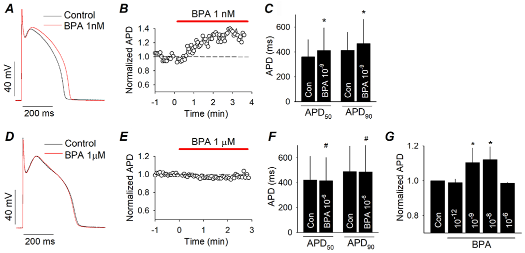
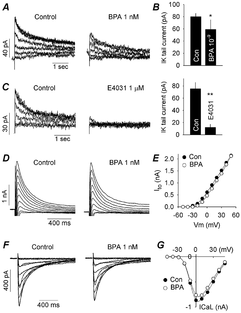
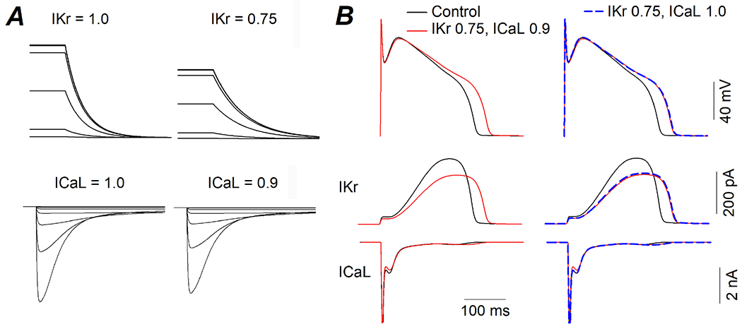
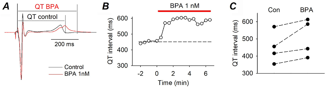
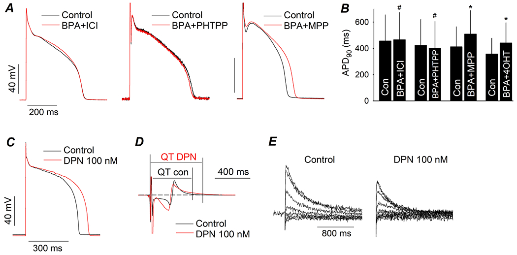
Bisphenol A (BPA) is a common environmental chemical with a range of potential adverse health effects. The impact of environmentally-relevant low dose of BPA on the electrical properties of the hearts of large animals (e.g., dog, human) is poorly defined. Perturbation of cardiac electrical properties is a key arrhythmogenic mechanism. In particular, delay of ventricular repolarization and prolongation of the QT interval of the electrocardiogram is a marker for the risk of malignant arrhythmias. We examined the acute effect of 10-9 M BPA on the electrical properties of female canine ventricular myocytes and tissues. BPA rapidly delayed action potential repolarization and prolonged action potential duration (APD). The dose response curve of BPA on APD was nonmonotonic. BPA rapidly inhibited the IKr K+ current and ICaL Ca2+ current. Computational modeling indicated that the effect of BPA on APD can be accounted for by its suppression of IKr. At the tissue level, BPA acutely prolonged the QT interval in 4 left ventricular wedges. ERβ signaling contributed to the acute effects of BPA on ventricular repolarization. Our results demonstrate that BPA has QT prolongation liability in female canine hearts. These findings have implication for the potential proarrhythmic cardiac toxicity of BPA in large animals.

Keywords: Arrhythmia marker; Bisphenol A; Canine heart; Low dose; QT prolongation; hERG channel.


Conflict of interest statement

Declaration of competing interest The authors declare that they have no known competing financial interests or personal relationships that could have appeared to influence the work reported in this paper.

Figures

Figure 1.
Figure 1.. Low dose BPA delays repolarization in female canine ventricular myocytes.
(A) Representative AP recorded from canine ventricular myocytes under control and upon exposure to 10−9 M BPA. (B) Time course of the action of 10−9 M BPA on APD. APD were normalized to the average value in control (dash line). (C) Average APD50 and APD90 under control and after acute exposure to 10−9 M BPA. N = 5 myocytes from 3 hearts. (D) Representative AP under control and upon exposure to 10−6 M BPA. (E) Time course of the action of 10−6 M BPA on APD. (F) Average APD50 and APD90 under control and after acute exposure to 10−6 M BPA. N = 4 myocytes from 2 hearts. (G) Average APD90 under control and upon acute exposure to BPA at indicated doses. N = 3 to 5 myocytes from 3 hearts. *: P < 0.05 vs control in a paired t-test or ANOVA. #: P > 0.1 vs control in a paired t-test. Error bars are S.D.
Figure 2.
Figure 2.. Acute impact of 10−9 M BPA on ionic currents in female canine ventricular myocytes.
(A) IKr tail current under control and upon rapid exposure to BPA. Tail currents were in response to depolarizing steps ranging from −20 to +30 mV in 10 mV increments from a holding potential of −40 mV, and recorded at −40 mV. (B) Average IKr tail current amplitude under control and after exposure to BPA. N = 5 myocytes from 5 hearts. (C) Effect of the selective IKr blocker E4031 on IKr tail current, confirming the identity of IKr. Right, average IKr tail current amplitude under control and in the presence of E4031. N = 3 myocytes from 2 hearts. (D) Representative Ito under control and upon exposure to BPA. Ito was activated with depolarizing steps ranging from - 30 to +50 mV in 10 mV increments, from a holding potential of −70mV at a frequency of 0.1 Hz. (E) Effect of BPA on Ito current-voltage relationship. (F) Representative ICaL under control and upon exposure to BPA. ICaL was elicited by voltage steps from −40 to +40 mV in 10 mV increment, from a holding potential of −50 mV at a frequency of 0.1 Hz. (G) Effect of BPA on ICaL current-voltage relationship. *: P < 0.05, **: P < 0.01 vs control in a paired t-test. Error bars are S.D.
Figure 3.
Figure 3.. Computational modeling of the effects of BPA on canine ventricular myocytes.
(A) Simulated voltage clamp experiments of Ikr (top) and ICaL (bottom) under control condition (current = 1.0) and simulated BPA exposure where IKr was reduced by a factor of 0.25 (25%) and ICaL was reduced by a factor of 0.1 (10%). (B) Action potentials (top) and concurrent IKr and ICaL currents (middle and bottom) that were elicited by the action potentials under indicated conditions.
Figure 4.
Figure 4.. Acute impact of 10−9 M BPA on QT interval in canine LV wedges.
(A) Representative EKG recorded from the same LV wedge under control and upon exposure to BPA. QT intervals are indicated. (B) Time course of the effect of BPA on QT interval. (C) QT intervals recorded from 4 LV wedges under control and upon exposure to 10−9 M BPA.
Figure 5.
Figure 5.. ERβ contributes to the effects of BPA on canine ventricular repolarization.
(A) Representative AP recorded from canine ventricular myocytes under control and upon exposure to 1 nM BPA in the presence of 1 μM ICI 182,780, 5 μM PHTPP, or 1 μM MPP. Myocytes were pretreated with the ER blocker for 15 min prior to exposure to BPA. (B) Average APD90 under control and upon exposure to BPA in the presence of ICI 182,780, PHTPP, MPP, or 1 μM 4-OHT. N = 3 to 4 myocytes from 1 to 3 hearts. (C to E), representative effect of 100 nM DPN on AP in ventricular myocytes, surface EKG from LV wedge, and IKr tail current. #: P > 0.1, *: P < 0.05 vs control in a paired t-test. Error bars are S.D.

Comment in

Similar articles

Cited by

References

    1. Abi-Gerges N, Philp K, Pollard C, Wakefield I, Hammond TG, Valentin JP, 2004. Sex differences in ventricular repolarization: from cardiac electrophysiology to Torsades de Pointes. Fundam Clin Pharmacol 18, 139–151. - PubMed
    1. Aboul Ezz HS, Khadrawy YA, Mourad IM, 2015. The effect of bisphenol A on some oxidative stress parameters and acetylcholinesterase activity in the heart of male albino rats. Cytotechnology 67, 145–155. - PMC - PubMed
    1. Arias-Loza PA, Jazbutyte V, Pelzer T, 2008. Genetic and pharmacologic strategies to determine the function of estrogen receptor alpha and estrogen receptor beta in cardiovascular system. Gend Med 5 Suppl A, S34–45. - PubMed
    1. Behere SP, Weindling SN, 2015. Inherited arrhythmias: The cardiac channelopathies. Ann Pediatr Cardiol 8, 210–220. - PMC - PubMed
    1. Belcher SM, Chen Y, Yan S, Wang HS, 2012. Rapid estrogen receptor-mediated mechanisms determine the sexually dimorphic sensitivity of ventricular myocytes to 17β-estradiol and the environmental endocrine disruptor bisphenol A. Endocrinology 153, 712–720. - PMC - PubMed