. 2023 Jan;9(1):142-156.
doi: 10.1038/s41477-022-01308-6. Epub 2022 Dec 30.

Cryo-EM structure of the respiratory I + III2 supercomplex from Arabidopsis thaliana at 2 Å resolution

Affiliations

Cryo-EM structure of the respiratory I + III2 supercomplex from Arabidopsis thaliana at 2 Å resolution

Niklas Klusch et al. Nat Plants. 2023 Jan.

Abstract

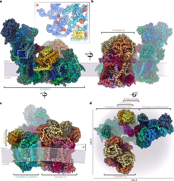
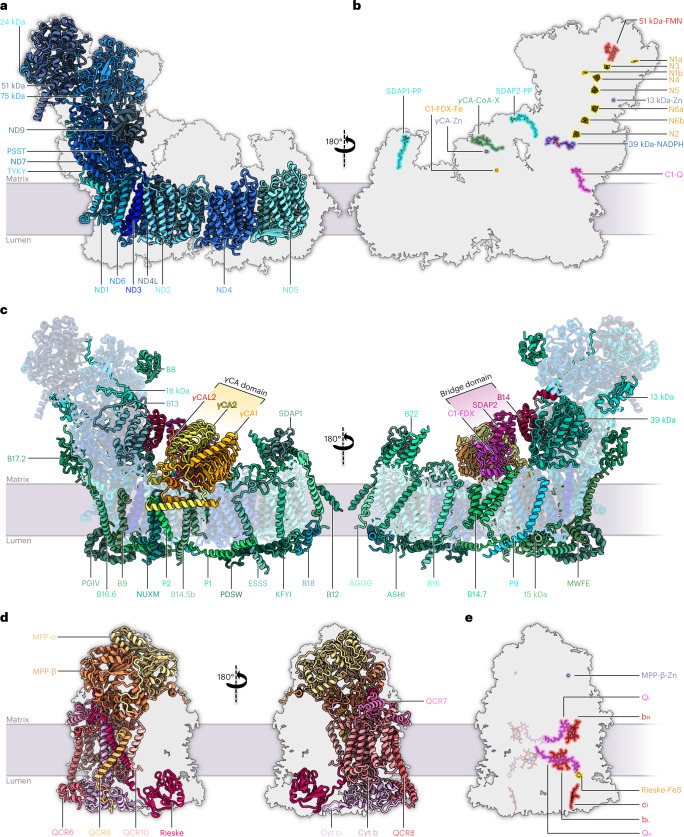
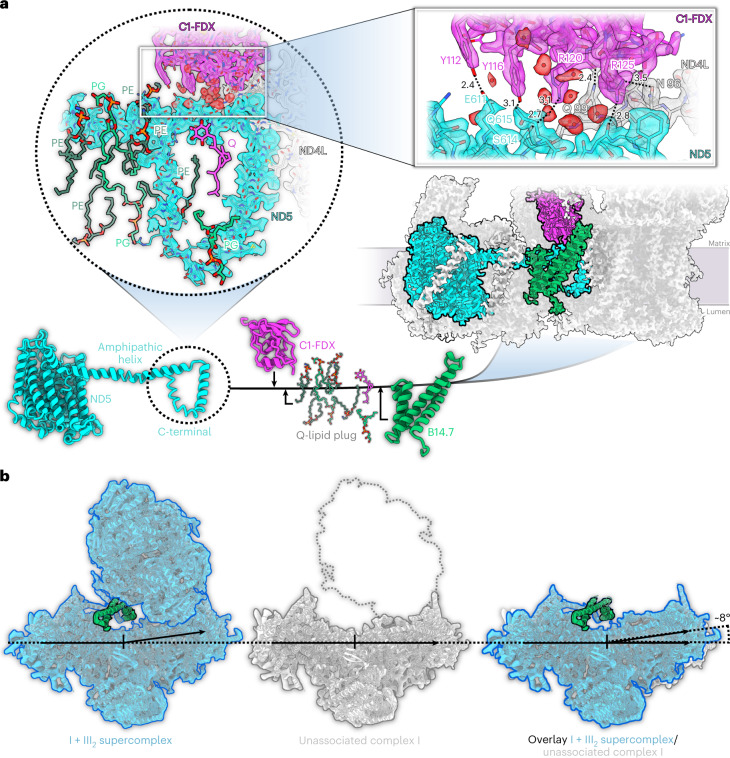
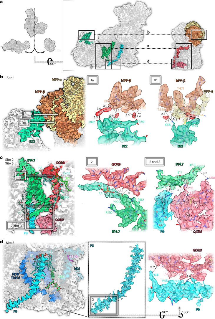
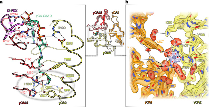
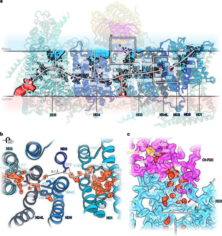
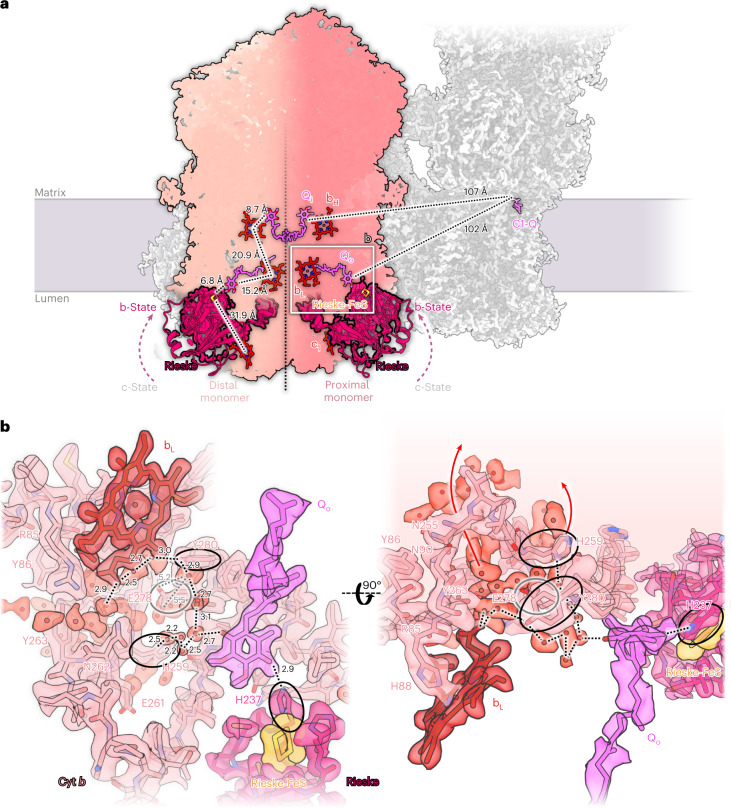
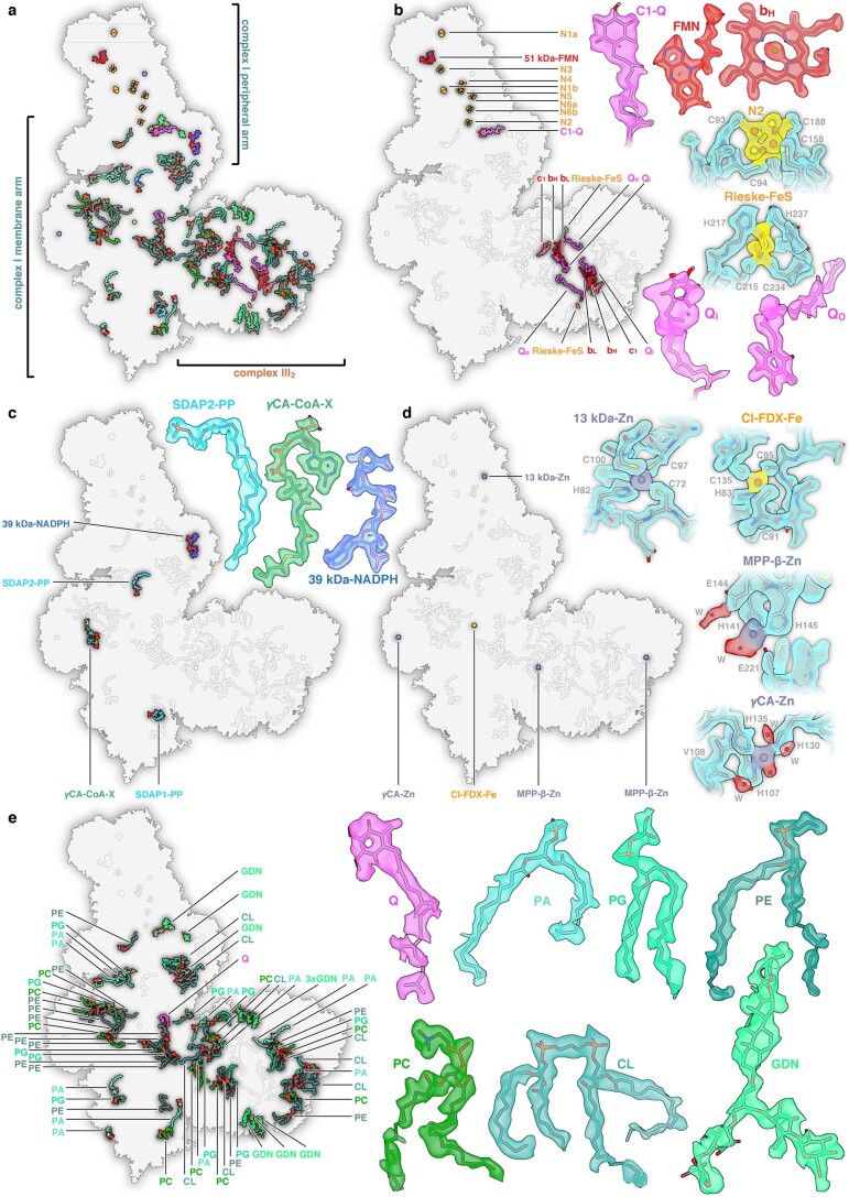
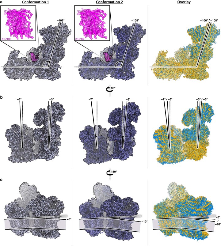
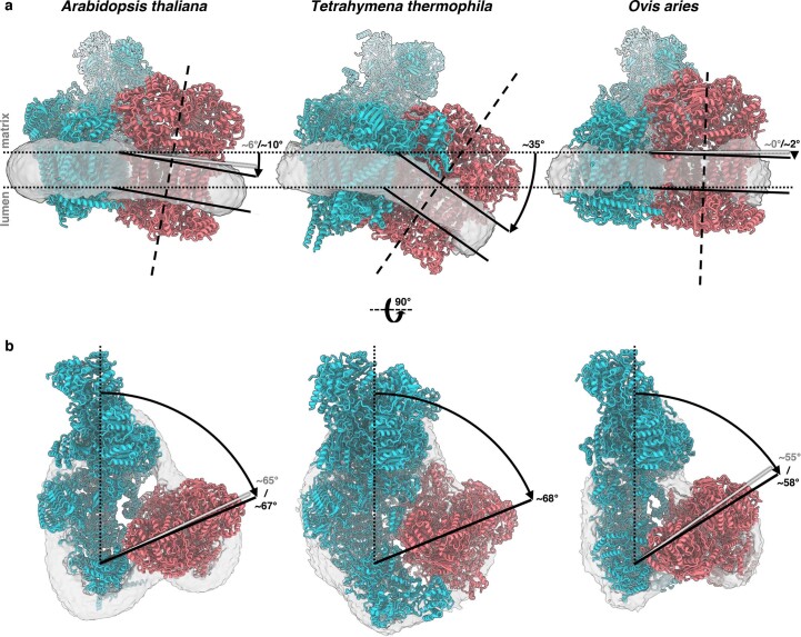
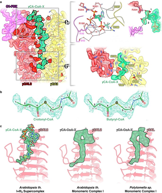
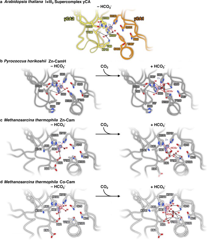
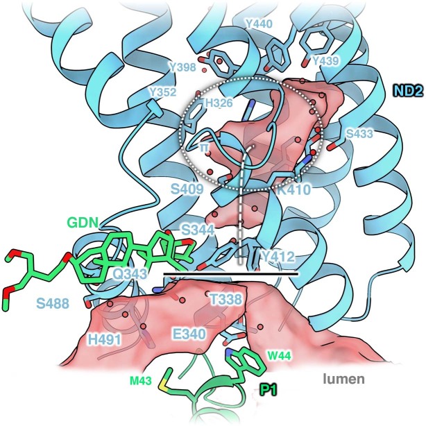
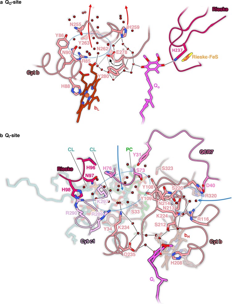
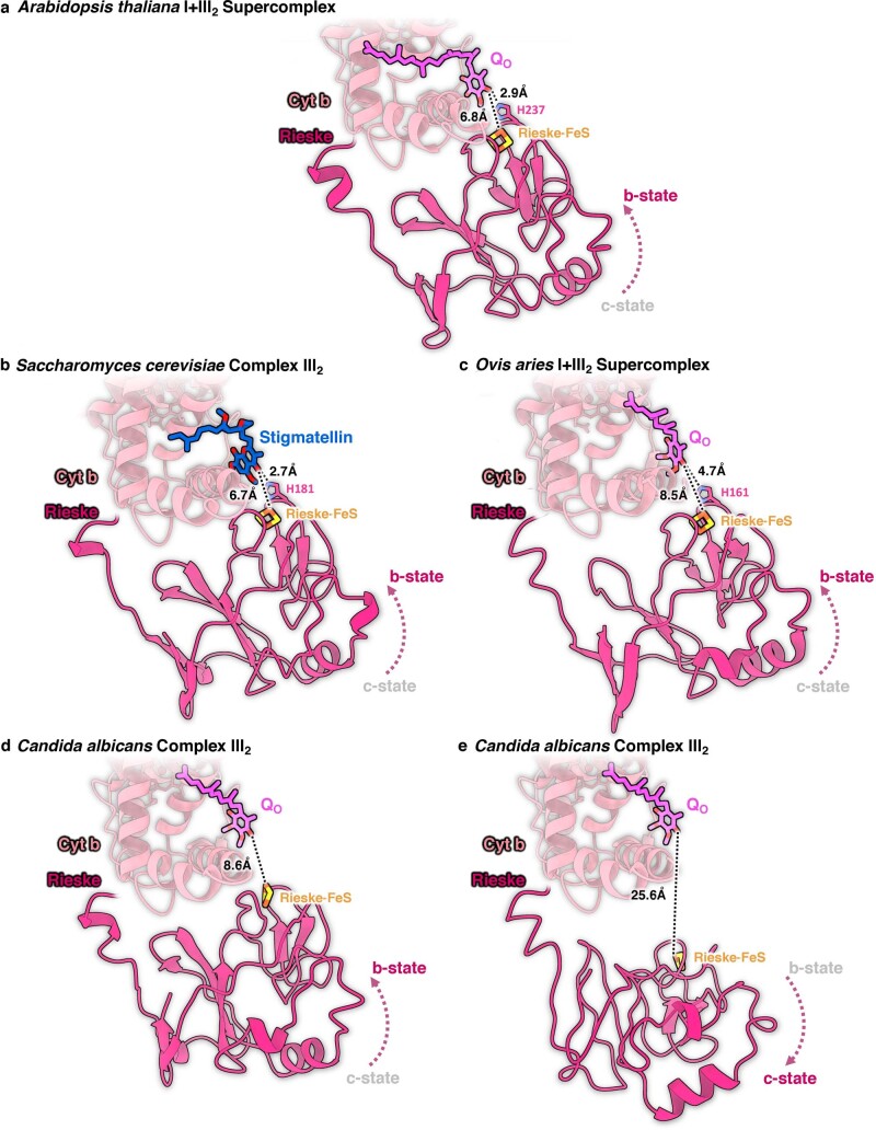
Protein complexes of the mitochondrial respiratory chain assemble into respiratory supercomplexes. Here we present the high-resolution electron cryo-microscopy structure of the Arabidopsis respiratory supercomplex consisting of complex I and a complex III dimer, with a total of 68 protein subunits and numerous bound cofactors. A complex I-ferredoxin, subunit B14.7 and P9, a newly defined subunit of plant complex I, mediate supercomplex formation. The component complexes stabilize one another, enabling new detailed insights into their structure. We describe (1) an interrupted aqueous passage for proton translocation in the membrane arm of complex I; (2) a new coenzyme A within the carbonic anhydrase module of plant complex I defining a second catalytic centre; and (3) the water structure at the proton exit pathway of complex III2 with a co-purified ubiquinone in the QO site. We propose that the main role of the plant supercomplex is to stabilize its components in the membrane.


Conflict of interest statement

The authors declare no competing interests.

Figures

Fig. 1
Fig. 1. 2 Å structure of the Arabidopsis I + III2 supercomplex.
ac, Views from the plane of the inner mitochondrial membrane (grey), with complex I in front (a), with complex III2 in front (b), and from the tip of the complex I membrane arm (c). The supercomplex protrudes into the mitochondrial matrix and the cristae lumen. The 14 complex I core subunits, which are conserved in bacterial and mitochondrial complex I, are drawn in shades of blue; accessory subunits in shades of green; the three subunits of the γCA module in yellow, orange and red; the three subunits of the bridge module in pink, purple and red. In a, the inset shows a typical map region near FeS cluster N2 (yellow), and water densities are red. In c, the inner mitochondrial membrane bends by ~8° around the supercomplex. d, Matrix view of the I + III2 supercomplex. Subunits protruding from the membrane are shown in strong colours. For detailed view of the high-resolution structure, see Supplementary Videos 1 and 2. For cryo-EM data processing and activity measurements, see Supplementary Figs. 2–5. All subunits are identified in Fig. 2. For comparison with the low-resolution map of the supercomplex obtained by single-particle negative-stain EM, see Supplementary Fig. 6.
Fig. 2
Fig. 2. Subunit composition of complex I and complex III2 within the Arabidopsis I + III2 supercomplex.
ac, Atomic model of complex I, showing the 14 core subunits in shades of blue (a), cofactors bound to complex I (N, FeS clusters; Q, ubiquinone/ubiquinol; FMN, flavine mononucleotide) (b) and accessory subunits of mitochondrial complex I (c). Conserved accessory subunits are shown in shades of green, the subunits of the carbonic anhydrase (CA) domain in yellow and orange, and the subunits of the bridge domain in red and pink. The newly identified subunit P9 is light blue. Subunit nomenclature as for bovine complex I (ref. 80) except for non-conserved accessory subunits (for details, see Supplementary Table 5). d,e, Atomic model of complex III2, showing the structure of the ten subunits of one complex III monomer within the complex III dimer (for details, see Supplementary Table 6) from opposite directions (d) and bound cofactors of complex III2 (bH, bL, c1: haem groups attached to cytochrome b and c1; FeS: iron–sulphur cluster attached to the Rieske protein; Qi, Qo: quinone binding sites; Zn: zinc2+ bound to MPP-β) (e). For details and bound lipids, see Extended Data Fig. 2.
Fig. 3
Fig. 3. The role of subunit B14.7 and C1-FDX in I + III2 supercomplex formation.
a, Subunit B14.7 (green) and C1-FDX (magenta) interact closely with the C-terminal loop of ND5 (cyan). The loop is surrounded by a set of lipids and a Q molecule (circled inset). C1-FDX sits on top of the amphipathic helix of ND5 and stabilizes it via a tight hydrogen bond network (black dotted lines) including well-defined water molecules (square inset; all distances in Å). b, Supercomplex (left, blue) with subunit B14.7 (green) compared with the structure of unassociated complex I (centre; grey), which does not have the B14.7 subunit. The position of the complex III dimer in the supercomplex is indicated by a grey dotted outline. An overlay (right) indicates that in the supercomplex the membrane arm of complex I rotates towards complex III2 by ~8°, resulting in a more extensive contact surface, which would stabilize the supercomplex. PE, phosphatidylethanolamine; PG, phosphatidylglycerol; Q, ubiquinone/ubiquinol.
Fig. 4
Fig. 4. Three interaction sites of complexes I and III2 in the Arabidopsis supercomplex.
a, Overview. b, Interaction site 1: B22 (green) of complex I binds to MPP-β (orange) and MPP-α (yellow) of complex III2. 1a and 1b show two roughly orthogonal views of interaction site 1. Interaction is mediated by hydrogen bonds including water molecules (red) and salt bridges. c, In site 2, B14.7 of complex I binds to QCR8 (red) and QCR6 (pink) of complex III2. In addition to polar contacts, the interaction is mediated by a set of membrane phospholipids. In site 3, binding to QCR6 involves the newly identified subunit P9 (light blue) of complex I. d, Also in site 3, the C-terminal part of P9 binds to QCR6 of complex III2 by salt bridges and hydrophobic contacts. Interacting amino acid residues are indicated by the one-letter code. Lipids: CL, cardiolipin; GDN, synthetic digitonin analogue glyco-diosgenin; PA, phosphatidic acid; PC, phosphatidylcholine; PE, phosphatidylethanolamine; PG, phosphatidyl-glycerol; Q, ubiquinone/ubiquinol. The network of hydrogen bonds (in Å) is indicated by black (<3.5 Å) or grey (>3.5 Å) dotted lines.
Fig. 5
Fig. 5. Catalytic sites in the γCA domain.
Overview of the γCA domain (centre) with γCA1 (orange), γCA2 (yellow) and γCAL2 (red). a, Catalytic site at the γCA2–γCAL2 interface with bound butyryl- or crotonyl-CoA, here referred to as γCA-CoA-X (green). Subunits γCA2 and γCAL2 interact with the phosphate groups and the adenine ring of CoA-X either directly or across a chain (black dotted lines) of water molecules (red). The 3′ phosphate group adopts two alternative conformations, as indicated by two strong densities in the cryo-EM map (Extended Data Fig. 5). The butyryl or crotonyl group of CoA-X is located at the putative catalytic site of the γCAL2–γCA2 interface (grey dotted ellipse). b, Map density and fitted atomic model of the catalytic site at the γCA1–γCA2 interface. A zinc ion is coordinated by three histidines and one water molecule in tetrahedral geometry (red dotted lines). The active site is surrounded by a network of hydrogen bonds. For details, see Extended Data Figs. 5 and 6.
Fig. 6
Fig. 6. Proton transfer path in the membrane arm of Arabidopsis complex I.
a, Conserved amino acids and water molecules (red spheres) in the membrane arm mark the central aqueous passage (white continuous line) and potential water channels (white dashed lines) to the mitochondrial matrix (transparent blue) or the cristae lumen (transparent red). The passage is interrupted at transmembrane helix 3 of subunit ND6 (red bars; see b). In subunit ND2, two potential half-channels connect the central aqueous passage to the matrix or the lumen. C1-FDX with its bound Fe ion sits next to the entrance of the ND2 matrix half-channel. The matrix and lumenal half-channel of ND5 are open. Proton leakage is prevented by a ~6 Å gap between ND5 H257 and T315 of the aqueous passage (red dotted line). b, π-Gate at the TMH3 of ND6 seen from the matrix side. Core subunits of the membrane arm are represented in shades of blue. The water chain in the central aqueous passage is interrupted by a ~9.1 Å gap. c, Density and fitted atomic model of the map region where C1-FDX (magenta) interacts with ND2 at the entrance to the potential aqueous half-channel that would connect the matrix to the central aqueous passage. The horizontal black line indicates that under our experimental conditions the channel is closed.
Fig. 7
Fig. 7. Mechanistic insights into Arabidopsis complex III2.
a, Overview of complex III cofactors involved in respiratory electron transport. Each monomer binds haems c1, bH, bL (red), a Rieske FeS cluster (orange) and a Q (magenta) at the reduction/oxidation site (Qi and Qo). In both monomers of complex III2, the Rieske head domain (dark red) is found in the b state. Complex III monomers proximal or distal to the ubiquinone binding site of complex I (C1-Q) are shown in different shades of pink divided by a black dotted line. Distances between the cofactors are shown for the distal complex III monomer. Distances from Qi and Qo to the quinol binding site in complex I (C1-Q) are shown for the proximal monomer. b, Detailed view of the proximal Qo site. Hydrogen network for release of the two protons during ubiquinol oxidation at the Qo site are shown by black dotted lines. One proton is transferred along a chain of water molecules (light red) via Cyt b Y280 and H259; the other can be transferred directly to H237 of the Rieske head domain. The three participating sidechains are indicated by black ellipses. Cyt b E278 (grey) may be involved in proton translocation, faces away from the bound native Q and is not part of a proton pathway (grey dotted lines). Red arrows indicate routes for proton release to the bulk solvent of the cristae lumen. For further details, see Extended Data Figs. 8 and 9.
Extended Data Fig. 1
Extended Data Fig. 1. Phylogenetic analysis of complex I subunit P9.
P9 is a two-helix (H1, H2) peptide that spans the inner mitochondrial membrane from the matrix to the lumenal side by its transmembrane segment (TMSEG). For phylogenetic analysis, Arabidopsis P9 (At1g67785) was used to search the non-redundant protein sequences (nr) database at the National Center for Biotechnology Information (NCBI, “https://www.ncbi.nlm.nih.gov/”) using standard settings. >100 homologous sequences were identified below an E-value of 1e-4. All sequences were from seed plants (spermatophyta). Sixteen sequences were selected for building a multiple sequence alignment using Clustal Omega (https://www.ebi.ac.uk/Tools/msa/clustalo/); five sequences for dicots and monocots, respectively, and all identified sequences of other spermatophyta clades. Amino acid positions conserved in 16/16 sequences are highlighted in dark blue, amino acid positions conserved in ≥14/16 sequences in mid-blue and amino acid positions conserved in ≥10/16 sequences in light blue. Phylogenetic clades are indicated to the left of the alignment. The topology model of P9 is indicated above the alignment. The model was built into the cryo-EM density of the Arabidopsis I + III2 supercomplex from valine at position 2 to alanine at position 56. The positions of its two alpha-helices are shown below the alignment. The complex III2 interacting segment is shown in red. Residues K41, V45, V48, R49, L52 of Arabidopsis P9 that interact directly with complex III2 are indicated by asterisks (see Fig. 4c and d). Accession of sequences. Arabidopsis thaliana: At1g67785, Brassica napus: CAF2186799, Medicago truncatula: XP_013446710, Malus domestica: XP_028959763, Trifolium pratense: XP_045824609, Pisum sativum: XP_050898094, Oryza sativa: NP_001392381, Hordeum vulgare: XP_044978029, Spirodela intermedia: CAA2619857, Phoenix dactylifera: XP_008797106, Asparagus officinalis: XP_020253321, Amborella trichopoda: XP_020527559, Nymphaea colorata: XP_031475842, Aristolochia fimbriata: KAG9445531, Picea sitchensis: ABK24944, Pinus taeda: AFG45619.
Extended Data Fig. 2
Extended Data Fig. 2. Bound cofactors, metal ions and lipids in the Arabidopsis I + III2 supercomplex.
a, Overview from the matrix side. b, Cofactors involved in respiratory electron transport. Complex I: FMN (Flavin mononucleotide), N1a, N1b, N2, N3, N4, N5, N6a, N6b (8 FeS clusters) and C1-Q (ubiquinol/ubiquinone at the Q reduction site). Complex III2: two copies each of heme bH, bL and c1, Rieske FeS, plus ubiquinone/ubiquinol at the Qo and Qi sites. c, Other cofactors: PP (S-acyl-4ʼ-phosphopantetheine bound to SDAP1 and SDAP2), CoA-X (Butyryl-or Crotonyl-Coenzym A bound to γCA), and NADPH bound to the 39 kDA subunit. d, Bound metal ions. e, Lipids and detergent molecules, with numbers of copies in round brackets. PA, phosphatidic acid (11); PC, phosphatidylcholine (9); PE, phosphatidylethanolamine (14); PG, phosphatidylglycerol (10); CL, cardiolipin (8); GDN, glyco-diosgenin (10); Q, ubiquinone/ubiquinol (not bound in the Q reduction site of complex I or in the Qo and Qi sites of complex III2). Densities and models of selected cofactors, metal ions and lipids are highlighted.
Extended Data Fig. 3
Extended Data Fig. 3. Conformations of the Arabidopsis I + III2 supercomplex.
Confromation 1 and 2 with their corresponding overlays on the right. a, View from the plane of the inner mitochondrial membrane indicating the angle between the peripheral and membrane arm of complex I in the two resolved, slightly different conformations of the I + III2 supercomplex. The angle is 106° in conformation 1 and 108° in confromation 2. The ferredoxin subunit of the bridge domain (C1-FDX) is present and well-defined in both conformations (magenta insets; complex III2 removed for clarity). b, An end-on view of the supercomplex shows that the peripheral arm tilts by 3 to 5° and C III2 by 3 to 7 ° relative to the membrane normal (dotted lines). c, A view from the opposite direction reveals that the membrane plane of complex III2 tilts by 6° or 10° relative to the membrane plane of complex I (dotted line). The panels on the right show conformation 1 in orange and conformation 2 in blue. The inner mitochondrial membrane is shown in grey. The two confromations were obtained by focused 3D classification (Supplementary Figs. 2 and 4; Supplementary Tables 1 and 2).
Extended Data Fig. 4
Extended Data Fig. 4. Conformations of the I + III2 supercomplex in different species.
Comparison of I + III2 supercomplex from Arabidopsis, Tetrahymena and ovine mitochondria. Atomic models of complex I are blue, complex III2 is red. The position of the inner mitochondrial membrane is indicated by the detergent micelles in the cryo-EM maps lowpass-filtered to 7 Å (grey). Left: conformation 2 of Arabidopsis thaliana; centre: consensus refinement of Tetrahymena thermophila; right: open conformation of Ovis aries. a, View from the tip of the complex I membrane arm, indicating the angle to which the membrane around complex III2 bends relative to complex I. The different angles in conformation 1 in Arabidopsis and the closed conformation of the ovine supercomplex are indicated by grey lines. b, Angle included between complex III2 and the complex I membrane arm as seen from the mitochondrial matrix. The dotted line indicates the approximate long axis of the complex I membrane; the straight black line is the transverse axis of complex III2. Grey lines indicate the transverse axis of Arabidopsis conformation 1 and the ovine closed conformation. The Arabidopsis structure is from this study, the structure of the open Ovis aries supercomplex is from (PDB: 6QC3, EMDB: 4495) and that of the Tetrahymena supercomplex from (PDB: 7TGH, EMDB: 25882).
Extended Data Fig. 5
Extended Data Fig. 5. Butyryl- or Crotonyl-CoA at the γCAL2/γCA2 interface in the Arabidopsis I + III2 supercomplex.
a, Left: Overview of atomic model and cryo-EM density at the complex I/III2 interface. γCAL2, light red; γCA2, yellow; C1-FDX, pink; water molecules, red densities or spheres; Butyryl- or Crotonyl-CoA (γCA-CoA-X), green. a, Right: Details of the CoA-X 3′-phosphate ADP binding region (top). The 3′-phosphate group can adopt two conformations as indicated by two strong densities in the cryo-EM map and interacts either with K40 of C1-FDX and R190 of γCAL2, or with R204 of γCAL2. γCAL2 N201 interacts either directly with the adenine ring or via a water molecule together with γCA2 S167. γCA2 R190 and a network of water molecules connect the diphosphate group of CoA-X to the three subunits at the interface. The butyryl or crotonyl group of CoA-X is located at the catalytic site of the γCAL2/γCA2 interface (below). The CoA-X oxygen of the C4 carbonyl group interacts via a water molecule with γCAL2 R152, γCAL2 H124, γCA2 H130 and γCA2 Y207. Q101 of subunit γCA2 is in direct hydrogen bond contact to the carbonyl group. Hydrogen bonds are indicated by dotted lines. b, Butyryl- or crotonyl-CoA fit the map density equally well. The model was drawn in Coot with the cryo-EM density at a contour level of 11.5 rmsd. c, A CoA-X density is also visible in the γCA domain of free complex I from Arabidopsis and Polytomella, but was not unambiguously identified at the lower map resolution.
Extended Data Fig. 6
Extended Data Fig. 6. Active site architecture of gamma-type carbonic anhydrases.
a, γCA-domain of complex I from Arabidopsis. The active site is located at the interface between the γCA2 (yellow) and γCA1 (orange) subunit. The three conserved histidines (H130 of γCA1, H107 and H135 of γCA2) coordinate a zinc ion (Zn) together with one water molecule (w) in a tetrahedral geometry (metal coordination shown by red dotted lines). The water molecule forms a hydrogen bond (black dotted lines) with an additional water that interacts with the polypeptide backbone of γCA2 V108 and residue γCA2 K110. A third water is coordinated by γCA2 D153. There appears to be no bound HCO3. Coordination of Zn, the hydrogen network and conserved amino acids including Q101, N99, Y207 and S203 resemble the active Zn-CamH site of Pyrococcus horikoshii. b, Pyrococcus horikoshii Zn-CamH. Left: without HCO3 (PDB: 1V3W), right: with HCO3 (PDB: 1V67). c, Methanosarcina thermophila Zn-Cam. Left: without HCO3 (PDB: 1QRG), right: with HCO3 (PDB: 1QRL). Note that the zinc ion at the Cam site is coordinated by two further water molecules compared to the γCA2/γCA1 site in Arabidopsis and CamH. In addition, E62 and E84 that are known to be important for proton release replace V89 and K110 of γCA2 and I47 and H68 of CamH. d, Methanosarcina thermophila Co-Cam. Left: without HCO3 (PDB: 1QQ0), right: with HCO3 (PDB: 1QRE). A cobalt ion (Co) is coordinated by the three conserved histidines and three additional water molecules. In contrast to the heterotrimeric CA domain of Arabidopsis complex I, the bacterial Cam/CamH enzymes are homotrimers with three identical active sites located at the three subunit interfaces.
Extended Data Fig. 7
Extended Data Fig. 7. A potential lumenal half-channel at core subunit ND2.
View along the membrane arm. In the Arabidopsis I + III2 supercomplex, a water-filled part of the central aqueous passage around the ND2 π-bulge (transparent red, white dotted ellipse) is blocked on the lumenal side (horizontal black line) by the sidechain of ND2 Y412 and an adjacent detergent molecule (GDN, green). Waters in red; GDN, glyco-diosgenin.
Extended Data Fig. 8
Extended Data Fig. 8. Architecture of the ubiquinol/ubiquinone (Q) binding sites in Arabidopsis complex III2.
a, The Qo site. One proton from bound ubiquinol can be transferred directly to H237 of the Rieske head domain in the b-state (black dotted line). The second proton can be released into the bulk solvent of the lumen (red arrows) along a chain of water molecules towards Cyt b H259 or towards a pool of water molecules (dotted blue ellipse) involving Cyt b Y280 and the heme bL propionate group. The sidechain of Cyt b E278 of the Qo motif faces away from the ubiquinol (black arrow) and its carboxylate group is not in hydrogen bond distance to the substrate or the water chain (grey dotted lines). b, The Qi site. Proton transfer from the bulk solvent of the matrix (blue arrows) towards a carbonyl group of bound ubiquinon can proceed via two hydrogen bond networks from clusters of water molecules (blue ellipses). One of them is surrounded by subunit QCR7, the N-terminal part of the Rieske protein, subunit Cyt c1, Cyt b and three bound lipids (two cardiolipins, CL; one phosphatidylcholine, PC). From this pool, protons can be accepted by Cyt b K234 from where they can pass to the bound ubiquinone across Cyt b D235. The other proton transfer pathway involves Cyt b K224. Our structure indicates that Cyt b H208 participates in proton transfer by forming a direct hydrogen bond to ubiquinone. Red dots, water molecules; black dotted lines, hydrogen bonds. Colors of the protein subunits (Cyt c1, Cyt b, Rieske, QCR7) as in Fig. 2. Q is drawn in pink.
Extended Data Fig. 9
Extended Data Fig. 9. Positions of the Rieske head domain in complex III structures.
In the Arabidopis I + III2 supercomplex (a), the Rieske head domain (magenta) is locked in the b-state with its FeS cluster positioned 6.8 Å and H237 2.9 Å away from the bound ubiquinone/ubiquinol that accepts the electron. These distances resemble those in the stigmatellin-bound complex III2 crystal structure of Saccharomyces cerevisiae (b) and are smaller than in the b-state in the cryo-EM structures of the Ovis aries (sheep) I + III2 supercomplex (c) and of the Candida albicans complex III2 (d) where the Rieske head group was also resolved in the c-state (e). Ubiquinone/ubiquinol is shown in pink, the inhibitor Stigmatellin in blue; distances between Qo/Rieske FeS group and Qo/FeS coordinating histidine are indicated by black dotted lines. PDB accession codes: Saccharomyces, 1EZV; Ovis, 6Q9E; Candida b-state, 7RJB, and Candida c-state, 7RJD. Sidechains of the Rieske head domain histidine are not present in the Candida models.

Comment in

  • Super-complex supercomplex.
    Møller IM. Møller IM. Nat Plants. 2023 Jan;9(1):5-6. doi: 10.1038/s41477-022-01329-1. Nat Plants. 2023. PMID: 36639556 No abstract available.

Similar articles

Cited by

References

    1. Hatefi Y, Haavik AG, Griffiths DE. Reconstitution of the electron transfer system I. Preparation and properties of the interacting enzyme complexes. Biochem. Biophys. Res. Commun. 1961;4:441–446. doi: 10.1016/0006-291X(61)90305-9. - DOI - PubMed
    1. Fowler LR, Richardson SH. Studies on the electron transfer system. L. On the mechanism of reconstitution of the mitochondrial electron transfer system. J. Biol. Chem. 1963;238:456–463. doi: 10.1016/S0021-9258(19)84019-X. - DOI - PubMed
    1. Hatefi Y, Rieske JS. The preparation and properties of DPNH-cytochrome c reductase (complex I–III of the respiratory chain) Methods Enzymol. 1967;10:225–231. doi: 10.1016/0076-6879(67)10044-X. - DOI
    1. Schägger H, Pfeiffer K. Supercomplexes in the respiratory chains of yeast and mammalian mitochondria. EMBO J. 2000;19:1777–1783. doi: 10.1093/emboj/19.8.1777. - DOI - PMC - PubMed
    1. Gu J, et al. The architecture of the mammalian respirasome. Nature. 2016;537:639–643. doi: 10.1038/nature19359. - DOI - PubMed

Publication types

MeSH terms

LinkOut - more resources