Review
. 2022 Dec 31;80(1):22.
doi: 10.1007/s00018-022-04670-y.

Vitreous humor proteome: unraveling the molecular mechanisms underlying proliferative and neovascular vitreoretinal diseases

Affiliations
Review

Vitreous humor proteome: unraveling the molecular mechanisms underlying proliferative and neovascular vitreoretinal diseases

Fátima Milhano Dos Santos et al. Cell Mol Life Sci. .

Abstract

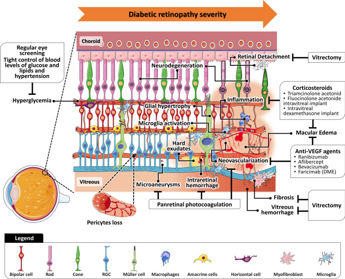
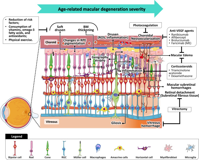
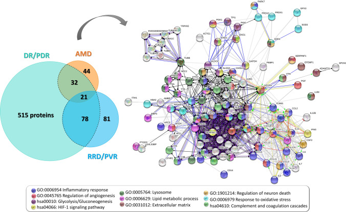
Proliferative diabetic retinopathy (PDR), proliferative vitreoretinopathy (PVR), and neovascular age-related macular degeneration (nAMD) are among the leading causes of blindness. Due to the multifactorial nature of these vitreoretinal diseases, omics approaches are essential for a deeper understanding of the pathophysiologic processes underlying the evolution to a proliferative or neovascular etiology, in which patients suffer from an abrupt loss of vision. For many years, it was thought that the function of the vitreous was merely structural, supporting and protecting the surrounding ocular tissues. Proteomics studies proved that vitreous is more complex and biologically active than initially thought, and its changes reflect the physiological and pathological state of the eye. The vitreous is the scenario of a complex interplay between inflammation, fibrosis, oxidative stress, neurodegeneration, and extracellular matrix remodeling. Vitreous proteome not only reflects the pathological events that occur in the retina, but the changes in the vitreous itself play a central role in the onset and progression of vitreoretinal diseases. Therefore, this review offers an overview of the studies on the vitreous proteome that could help to elucidate some of the pathological mechanisms underlying proliferative and/or neovascular vitreoretinal diseases and to find new potential pharmaceutical targets.

Keywords: Age-related macular degeneration; Proliferative diabetic retinopathy; Proliferative vitreoretinopathy; Vitreous proteomics.


Conflict of interest statement

The authors declare that they have no competing interests.

Figures

Fig. 1
Fig. 1
Vitreous and retinal anatomy in pathophysiological events in diabetic retinopathy and proliferative diabetic retinopathy and current treatments for each clinical feature. RGC retinal ganglion cell
Fig. 2
Fig. 2
Vitreous and retinal anatomy in pathophysiological events in the dry age-related macular degeneration and neovascular age-related macular degeneration and current treatments for each clinical feature. BM Bruch’s membrane, RGC retinal ganglion cell, ROS reactive oxygen species, oxidative stress, RPE retinal pigment epithelium
Fig. 3
Fig. 3
Vitreous and retinal anatomy in pathophysiological events related to the progression from retinal detachment to proliferative vitreoretinopathy and current treatments for each clinical feature. The experimental treatments are marked with *. ECM extracellular matrix, iBRB inner blood-retinal barrier, oBRB outer blood-retinal barrier, RGC retinal ganglion cell, RPE retinal pigment epithelium
Fig. 4
Fig. 4
Proteins detected in proteomics and multiplex studies as differentially expressed in the vitreous of patients with non- and proliferative diabetic retinopathy (DR/PDR), age-related macular degeneration (AMD), and reghmatogeneous retinal detachment (RRD)/proliferative vitreoretinopathy are depicted in the Venn diagram. Protein–protein interaction network of the proteins found to be differentially expressed in more than one disease (black dashed line in Venn diagram) obtained with high confidence in STRING v11.5. Nodes corresponding to the top enriched terms of Gene Ontology and KEGG pathways are highlighted in different colors, as shown in the caption of the figure. Disconnected nodes were removed from the protein–protein interaction network

Similar articles

Cited by

References

    1. Sabanayagam C, Cheng C-Y. Global causes of vision loss in 2015: are we on track to achieve the Vision 2020 target? Lancet Glob Heal. 2017;5:e1164–e1165. doi: 10.1016/S2214-109X(17)30412-6. - DOI - PubMed
    1. Bourne RRA, Flaxman SR, Braithwaite T, et al. Magnitude, temporal trends, and projections of the global prevalence of blindness and distance and near vision impairment: a systematic review and meta-analysis. Lancet Glob Heal. 2017;5:e888–e897. doi: 10.1016/S2214-109X(17)30293-0. - DOI - PubMed
    1. Ackland P, Resnikoff S, Bourne R. World blindness and visual impairment: despite many successes, the problem is growing. Community Eye Heal. 2018;30:71–73. - PMC - PubMed
    1. Swenor BK, Ehrlich JR. Comment Ageing and vision loss: looking to the future. Lancet. 2021 doi: 10.1016/S2214-109X(21)00031-0. - DOI - PubMed
    1. Yoshida S, Nakama T, Ishikawa K, et al. Periostin in vitreoretinal diseases. Cell Mol Life Sci. 2017;74:4329–4337. doi: 10.1007/s00018-017-2651-5. - DOI - PMC - PubMed

MeSH terms