

Posterior white matter hyperintensities are associated with reduced medial temporal lobe subregional integrity and long-term memory in older adults
- PMID: 36586358
- PMCID: PMC9830310
- DOI: 10.1016/j.nicl.2022.103308
Posterior white matter hyperintensities are associated with reduced medial temporal lobe subregional integrity and long-term memory in older adults
Abstract
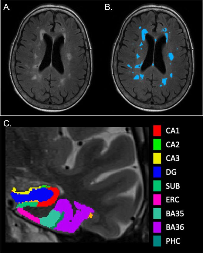
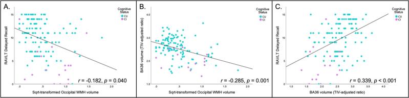
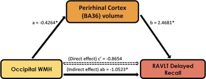
White matter hyperintensities are a marker of small vessel cerebrovascular disease that are strongly related to cognition in older adults. Similarly, medial temporal lobe atrophy is well-documented in aging and Alzheimer's disease and is associated with memory decline. Here, we assessed the relationship between lobar white matter hyperintensities, medial temporal lobe subregional volumes, and hippocampal memory in older adults. We collected MRI scans in a sample of 139 older adults without dementia (88 females, mean age (SD) = 76.95 (10.61)). Participants were administered the Rey Auditory Verbal Learning Test (RAVLT). Regression analyses tested for associations among medial temporal lobe subregional volumes, regional white matter hyperintensities and memory, while adjusting for age, sex, and education and correcting for multiple comparisons. Increased occipital white matter hyperintensities were related to worse RAVLT delayed recall performance, and to reduced CA1, dentate gyrus, perirhinal cortex (Brodmann area 36), and parahippocampal cortex volumes. These medial temporal lobe subregional volumes were related to delayed recall performance. The association of occipital white matter hyperintensities with delayed recall performance was fully mediated statistically only by perirhinal cortex volume. These results suggest that white matter hyperintensities may be associated with memory decline through their impact on medial temporal lobe atrophy. These findings provide new insights into the role of vascular pathologies in memory loss in older adults and suggest that future studies should further examine the neural mechanisms of these relationships in longitudinal samples.
Keywords: Aging; Cerebrovascular disease; Hippocampus; Long-term memory; Small vessel disease.
Copyright © 2022. Published by Elsevier Inc.
Conflict of interest statement
Declaration of Competing Interest The authors declare that they have no known competing financial interests or personal relationships that could have appeared to influence the work reported in this paper.
Figures
References
-
- Andersson C., Lindau M., Almkvist O., Engfeldt P., Johansson S.-E., Jönhagen M.E. Identifying patients at high and low risk of cognitive decline using Rey Auditory Verbal Learning Test among middle-aged memory clinic outpatients. Dement. Geriatr. Cogn. Disord. 2006;21(4):251–259. - PubMed
-
- Braak H., Braak E. Neuropathological stageing of Alzheimer-related changes. Acta Neuropathol. 1991;82(4):239–259. - PubMed
Publication types
MeSH terms
Grants and funding
LinkOut - more resources
Full Text Sources
Medical
Miscellaneous
