. 2023 Jan 31;42(1):111903.
doi: 10.1016/j.celrep.2022.111903. Epub 2022 Dec 14.

A delicate balance between antibody evasion and ACE2 affinity for Omicron BA.2.75

Affiliations

A delicate balance between antibody evasion and ACE2 affinity for Omicron BA.2.75

Jiandong Huo et al. Cell Rep. .

Abstract

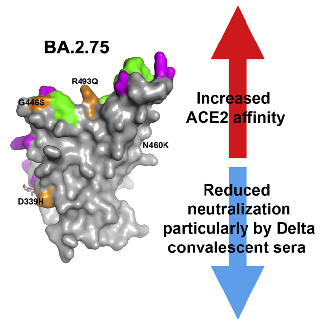
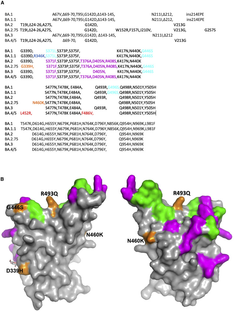
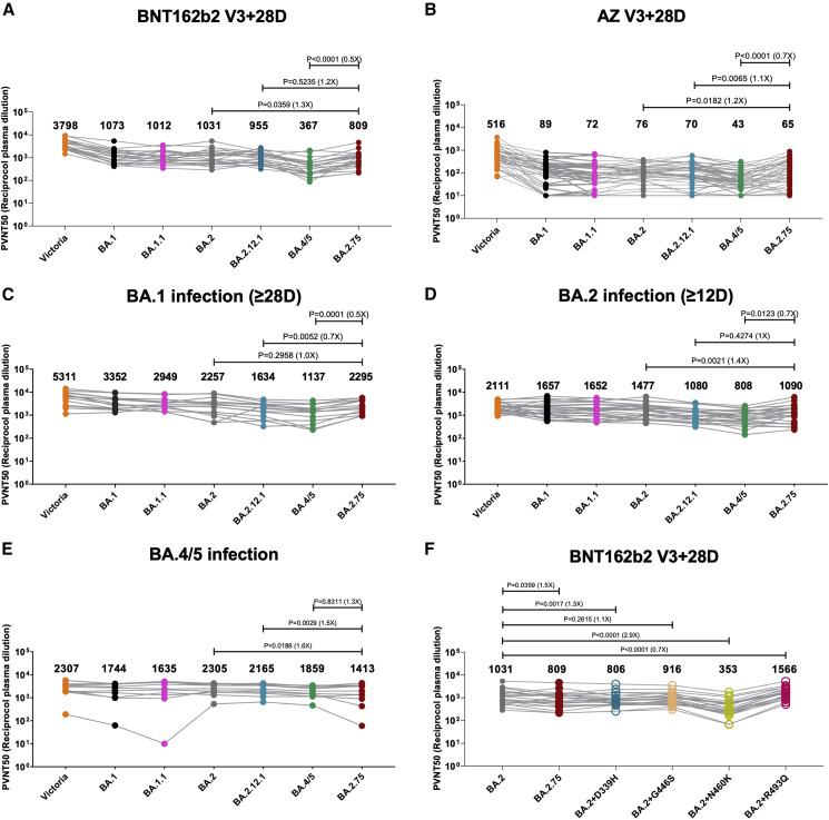
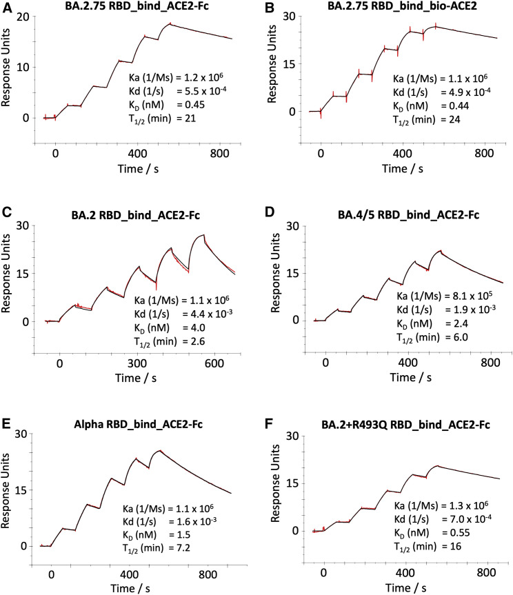
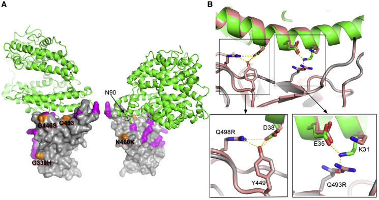
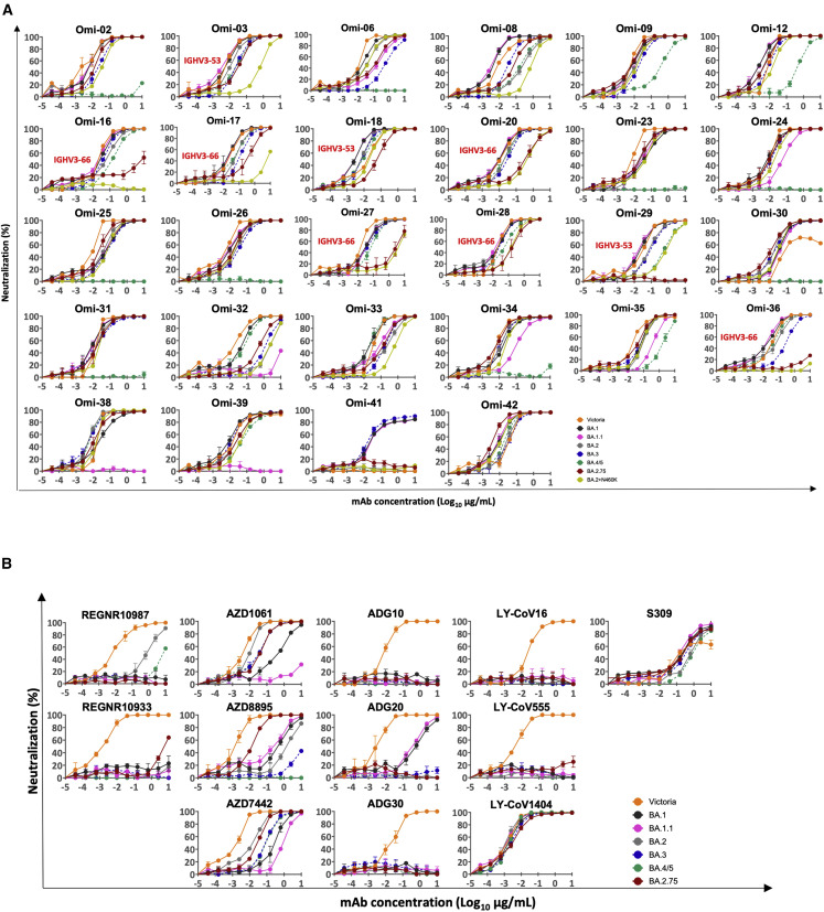
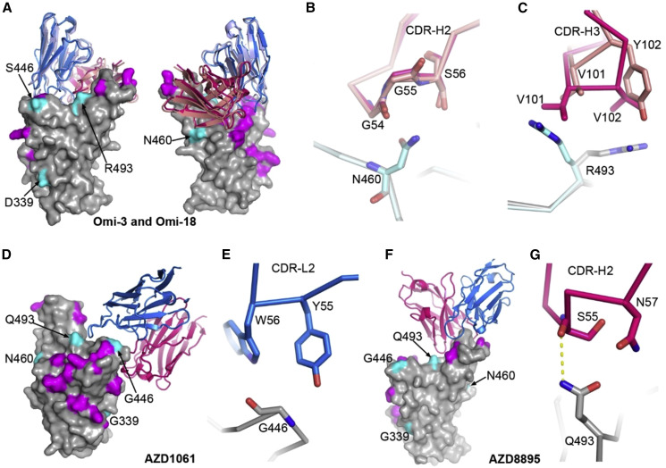
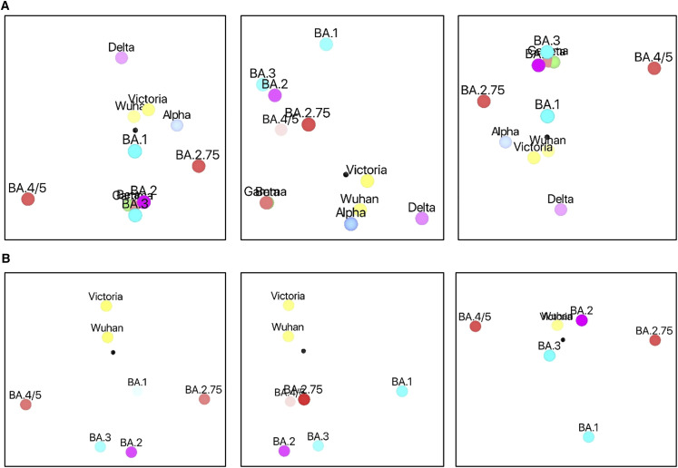
Variants of severe acute respiratory syndrome coronavirus 2 (SARS-CoV-2) have caused successive global waves of infection. These variants, with multiple mutations in the spike protein, are thought to facilitate escape from natural and vaccine-induced immunity and often increase in affinity for ACE2. The latest variant to cause concern is BA.2.75, identified in India where it is now the dominant strain, with evidence of wider dissemination. BA.2.75 is derived from BA.2 and contains four additional mutations in the receptor-binding domain (RBD). Here, we perform an antigenic and biophysical characterization of BA.2.75, revealing an interesting balance between humoral evasion and ACE2 receptor affinity. ACE2 affinity for BA.2.75 is increased 9-fold compared with BA.2; there is also evidence of escape of BA.2.75 from immune serum, particularly that induced by Delta infection, which may explain the rapid spread in India, where where there is a high background of Delta infection. ACE2 affinity appears to be prioritized over greater escape.

Keywords: ACE2 receptor; BA.2.75; COVID-19; CP: Immunology; CP: Microbiology; RBD; SARS-CoV-2; antigenic variation; immune escape; spike; variant; variant of concern.


Conflict of interest statement

Declaration of interests G.R.S. sits on the GSK Vaccines Scientific Advisory Board, consults for Astra Zeneca, and is a founding member of RQ Biotechnology. Oxford University holds intellectual property related to the Oxford-Astra Zeneca vaccine and SARS-CoV-2 mAbs discovered in G.R.S.’s laboratory. A.J.P. is Chair of UK Dept. Health and Social Care’s (DHSC) Joint Committee on Vaccination & Immunisation (JCVI) but does not participate in the JCVI COVID-19 committee and is a member of the WHO’s SAGE. The views expressed in this article do not necessarily represent the views of DHSC, JCVI, or WHO. The University of Oxford has entered into a partnership with AstraZeneca on coronavirus vaccine development. T.L. is named as an inventor on a patent application covering this SARS-CoV-2 vaccine and was a consultant to Vaccitech for an unrelated project whilst the study was conducted. S.J.D. is a scientific advisor to the Scottish Parliament on COVID-19.

Figures

None
Graphical abstract
Figure 1
Figure 1
Sequence changes in BA.2.75 compared with other Omicron sublineages (A) Sequence alignments of BA.2.75 together with Omicron sublineages Omicron BA.1, BA.1.1, BA.2, BA.3, and BA.4/5 boundaries of the NTD and RBD are marked. (B) Surface representation of mutated residues in the BA.2.75 RBD compared with the BA.2 RBD. Positions of BA.2 RBD mutations (gray surface with the ACE2 footprint in dark green) are shown, and residues mutated in BA.2.75 are shown in orange and labeled.
Figure 2
Figure 2
Pseudoviral neutralization assays of BA.2.75 by vaccine and BA.1, BA.2, and BA.4/5 immune serum (A and B) IC50 values for the indicated viruses using serum obtained from vaccinees 28 days following their third dose of vaccine (A) Pfizer BNT162b2 (n = 22) or (B) AstraZeneca AZD AZD1222 (n = 41). (C–E) Serum from volunteers suffering vaccine breakthrough BA.1 (n = 16), BA.2 (n = 23), or BA.4/5 (n = 11) infections. (F) IC50 values for single RBD point mutations inserted into the BA.2 pseudovirus using Pfizer BNT162b2 serum (n = 22). Geometric mean titers are shown above each column. The Wilcoxon matched-pairs signed rank test was used for the analysis and two-tailed p values were calculated. See also Table S3.
Figure 3
Figure 3
ACE2/RBD affinity SPR sensorgrams showing ACE2 binding of BA.2.75 RBD using ACE2-Fc (A) or biotinylated ACE2 as ligand (B) compared with binding to the RBD of BA.2 (C), BA.4/5 (D), Alpha (E), and BA.2 + R493Q (F). The data for BA.2, BA.4/5, and Alpha have been reported previously in Tuekprakhon et al., Dejnirattisai et al., and Nutalai et al., respectively.
Figure 4
Figure 4
The structure of BA.2.75 RBD/ACE2 complex (A) Front and back views of the overall structure of the BA.2.75 RBD/ACE2 complex. ACE2 is shown as green ribbons and the RBD as surface with mutations common to BA.2 highlighted in magenta and those that are different in orange. (B) BA.2.75 RBD (gray) and ACE2 (green) interface compared with that of BA.2 and ACE2 (both in salmon). Close ups show interactions of Q498R and Q493 (R493 in BA.2) with ACE2. See also Table S5.
Figure 5
Figure 5
Pseudoviral neutralization assays against monoclonal antibodies (A) Neutralization curves for a panel of 28 mAbs made from samples taken from vaccinees infected with BA.1. Titration curves for BA.2.75 are compared with Victoria, BA.1, BA.1.1, BA.2, and BA.4/5. IC50 titers are shown in Table S1A. (B) Pseudoviral neutralization assays with mAbs developed for human use. IC50 titers are shown in Table S1B. Data for Victoria, BA.1, BA.1.1, BA.2, and BA.4/5 are used for comparison and are taken from Tuekprakhon et al. See also Figure S1. All assays have been done at least twice.
Figure 6
Figure 6
Interactions between mAbs and BA.2.75 mutation sites (A) Front and back views of the binding modes of Omi-3 (PDB: 7ZF3) and Omi-18 (PDB: 7ZFC) complexed with Omicron BA.1 RBD by overlapping the RBD. The RBD is shown as a gray surface representation with mutations common to both BA.2 and BA.2.75 colored in magenta, and the four mutations that differ between the two are in cyan. Vhs and Vls are shown as ribbons and colored in red and blue for Omi-3 and light blue and salmon for Omi-18, respectively. (B) Interactions between N460 of the RBD and CDR-H2 of the Fabs. (C) Contacts between R493 of the RBD and CDR-H3 of the Fabs. In (B and C), the RBD associated with Omi-3 is in gray and Omi-18 in cyan, and the colors of the Fabs are as in (A). (D and E) AZD1061 bound with the ancestral SARS-CoV-2 RBD (PDB: 7L7E) (D) and contacts between G446 of the RBD and CDR-L2 of the Fab (E). (F and G) AZD8895 bound with the ancestral SARS-CoV-2 spike RBD (PDB: 7L7E) (F) and contacts between Q493 of the RBD and CDR-H2 of the Fab (G). In (D–F), the RBD is drawn and colored as in (A), and heavy chain is in red and light chain in blue.
Figure 7
Figure 7
Antigenic mapping (A) Orthogonal views of the antigenic map showing BA.2.75 in the context of the positions of early pandemic viruses, previous VoCs, and Omicron sublineages calculated from pseudovirus neutralization data. Distance between two positions is proportional to the reduction in neutralization titer when one of the corresponding strains is challenged with serum derived by infection by the other. No scale is provided since the figures are projections of a 3D distribution; however, the variation can be calibrated by comparison with (1) BA.1 to BA.2, which is 2.93× reduced, and (2) BA.2 to BA.4/5, which is 3.03× reduced. (B) As (A) but including only Omicron sublineages and early pandemic viruses to allow more accurate projection of this subset into three dimensions. Note that responses of these viruses against all sera were included in the calculations. See also Table S1.

References

    1. Robson F., Khan K.S., Le T.K., Paris C., Demirbag S., Barfuss P., Rocchi P., Ng W.L. Coronavirus RNA proofreading: molecular basis and therapeutic targeting. Mol. Cell. 2020;79:710–727. - PMC - PubMed
    1. Greninger A.L., Dien Bard J., Colgrove R.C., Graf E.H., Hanson K.E., Hayden M.K., Humphries R.M., Lowe C.F., Miller M.B., Pillai D.R., et al. Clinical and infection prevention applications of severe acute respiratory syndrome coronavirus 2 genotyping: an infectious diseases society of America/American society for microbiology consensus review document. J. Clin. Microbiol. 2022;60:e0165921. - PMC - PubMed
    1. Obermeyer F., Jankowiak M., Barkas N., Schaffner S.F., Pyle J.D., Yurkovetskiy L., Bosso M., Park D.J., Babadi M., MacInnis B.L., et al. Analysis of 6.4 million SARS-CoV-2 genomes identifies mutations associated with fitness. Science. 2022;376:1327–1332. - PMC - PubMed
    1. Sadeghalvad M., Mansourabadi A.H., Noori M., Nejadghaderi S.A., Masoomikarimi M., Alimohammadi M., Rezaei N. Recent developments in SARS-CoV-2 vaccines: a systematic review of the current studies. Rev. Med. Virol. 2022:e2359. - PMC - PubMed
    1. Focosi D., McConnell S., Casadevall A., Cappello E., Valdiserra G., Tuccori M. Monoclonal antibody therapies against SARS-CoV-2. Lancet Infect. Dis. 2022;22:e311–e326. - PMC - PubMed

Publication types