. 2023 Feb;299(2):102845.
doi: 10.1016/j.jbc.2022.102845. Epub 2022 Dec 28.

Targeting peroxisomal fatty acid oxidation improves hepatic steatosis and insulin resistance in obese mice

Affiliations

Targeting peroxisomal fatty acid oxidation improves hepatic steatosis and insulin resistance in obese mice

Haoya Yao et al. J Biol Chem. 2023 Feb.

Abstract

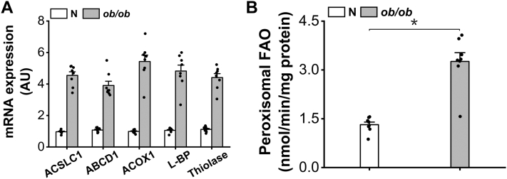
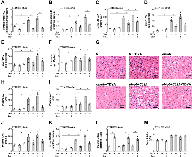
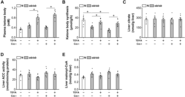
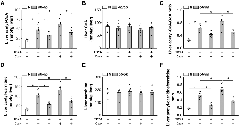
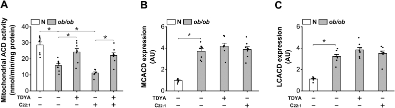
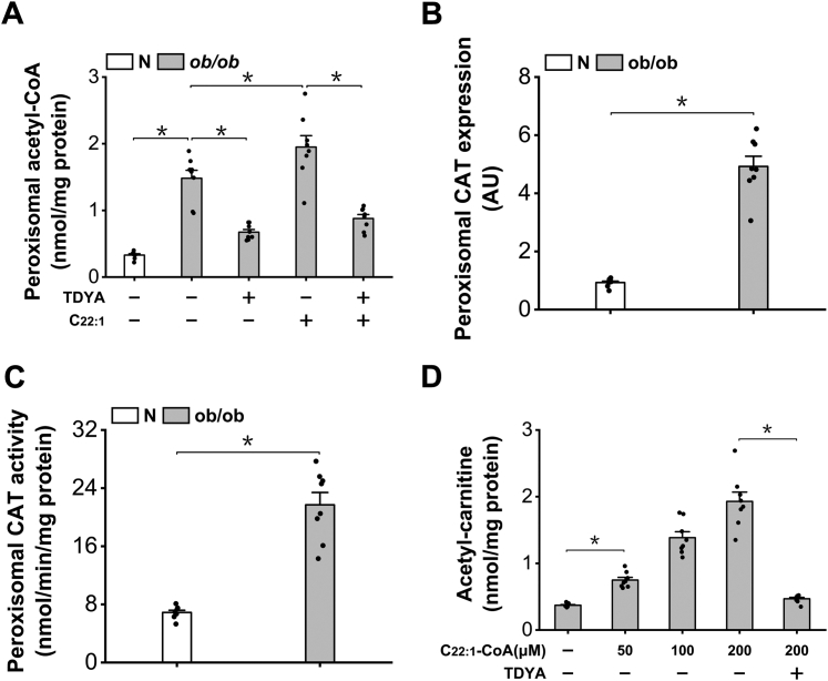
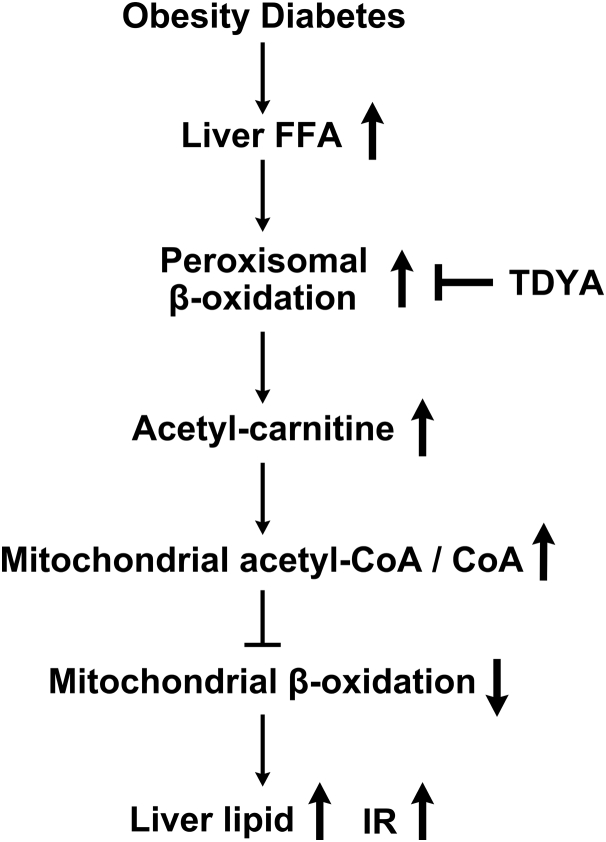
Obesity and diabetes normally cause mitochondrial dysfunction and hepatic lipid accumulation, while fatty acid synthesis is suppressed and malonyl-CoA is depleted in the liver of severe obese or diabetic animals. Therefore, a negative regulatory mechanism might work for the control of mitochondrial fatty acid metabolism that is independent of malonyl-CoA in the diabetic animals. As mitochondrial β-oxidation is controlled by the acetyl-CoA/CoA ratio, and the acetyl-CoA generated in peroxisomal β-oxidation could be transported into mitochondria via carnitine shuttles, we hypothesize that peroxisomal β-oxidation might play a role in regulating mitochondrial fatty acid oxidation and inducing hepatic steatosis under the condition of obesity or diabetes. This study reveals a novel mechanism by which peroxisomal β-oxidation controls mitochondrial fatty acid oxidation in diabetic animals. We determined that excessive oxidation of fatty acids by peroxisomes generates considerable acetyl-carnitine in the liver of diabetic mice, which significantly elevates the mitochondrial acetyl-CoA/CoA ratio and causes feedback suppression of mitochondrial β-oxidation. Additionally, we found that specific suppression of peroxisomal β-oxidation enhances mitochondrial fatty acid oxidation by reducing acetyl-carnitine formation in the liver of obese mice. In conclusion, we suggest that induction of peroxisomal fatty acid oxidation serves as a mechanism for diabetes-induced hepatic lipid accumulation. Targeting peroxisomal β-oxidation might be a promising pathway in improving hepatic steatosis and insulin resistance as induced by obesity or diabetes.

Keywords: acetyl-carnitine; fatty acid oxidation; mitochondria; obesity; peroxisomes.


Conflict of interest statement

Conflict of interest The authors declare that they have no conflicts of interest with the contents of this article.

Figures

Figure 1
Figure 1
Peroxisomal β-oxidation is induced in the liver of ob/ob mice.A, gene expressions of the enzymes involved in peroxisomal β-oxidation were induced in the liver of ob/ob mice. B, peroxisomal β-oxidation increased remarkably in the liver of ob/ob mice. ∗p < 0.05 by t test between paired groups.
Figure 2
Figure 2
Suppression of peroxisomal β-oxidation improves hepatic steatosis in ob/ob mice.A, peroxisomal β-oxidation in the liver of ob/ob mice was suppressed after treatment with TDYA. B, TDYA treatment significantly reduced hydrogen peroxide formation in the liver of ob/ob mice. C, LC-CoA increased significantly in the liver of ob/ob mice, which was reduced by the treatment of TDYA. D, liver TAG was significantly higher in the liver of ob/ob mice, as further increased by the treatment of C22:1 and reduced by TDYA. E, liver DAG was significantly higher in the liver of ob/ob mice, as further increased by the treatment of C22:1 and reduced by TDYA. F, C22:1 treatment significantly elevated the liver ratio of ob/ob mice, as lowered by the treatment of TDYA. G, liver histologic changes in ob/ob mice treated with C22:1 and TDYA. H and I, plasma (H) ALT and (I) AST were elevated remarkably in ob/ob mice compared to the normal group, as lowered by the treatment of TDYA. J, plasma TAG was significantly higher in ob/ob mice than in normal group, as reduced by the treatment with TDYA. K, TBARS increased significantly in the liver of ob/ob mice, as further increased by the treatment of C22:1 and reduced by TDYA. L, TDYA treatment caused significant elevation in plasma C26:0 in ob/ob mice. M, daily food intake was not affected in ob/ob mice treated with TDYA or C22:1. ∗p < 0.05 by t test between paired groups. TDYA, 10,12-tricosadiynoic acid; LC-CoA, long-chain acyl-CoA; TAG, triacylglyceride; DAG, diacylglyceride; ALT, alanine aminotransferase; AST, aspartate aminotransferase; TBARS, Thiobarbituric acid-reactive substances.
Figure 3
Figure 3
Suppression of peroxisomal β-oxidation improves insulin resistance in ob/ob mice.A, TDYA treatment significantly lowered plasma glucose of ob/ob mice. B, OGTT was improved in ob/ob mice treated with TDYA. C, plasma insulin increased remarkably in ob/ob mice, as lowered after treatment with TDYA. D, HOMA-IR was significantly higher in ob/ob mice, as further enhanced by the treatment of C22:1 and lowered by TDYA. ∗p < 0.05 by t test between paired groups. TDYA, 10,12-tricosadiynoic acid; IR, insulin resistance; HOMA-IR, homeostasis model assessment of insulin resistance; OGTT, oral glucose tolerance test.
Figure 4
Figure 4
Induction of peroxisomal β-oxidation suppresses mitochondrial FAO independent of malonyl-CoA.A, C22:1 feeding lowered and TDYA treatment elevated plasma ketone body in ob/ob mice. B, ketone body synthesis rate was significantly lower in ob/ob mice than the normal group, as further decreased by C22:1 feeding and recovered by TDYA. C, liver citrate content was not altered significantly among all the groups. D, liver ACC activity was not altered among all the groups. E, liver malonyl-CoA was not significantly changed in ob/ob mice treated with C22:1 or TDYA. ∗p < 0.05 by t test between paired groups. FAO, fatty acid oxidation; TDYA, 10,12-tricosadiynoic acid; ACC, acetyl-CoA carboxylase.
Figure 5
Figure 5
Peroxisomal β-oxidation of fatty acids increases mitochondrial acetyl-CoA/CoA ratio.A, liver acetyl-CoA increased considerably in the liver of ob/ob mice, and C22:1 feeding caused further increase in mitochondrial acetyl-CoA, which was lowered by TDYA. B, liver free CoA was not altered significantly among all the groups. C, liver acetyl-CoA/CoA ratio was elevated significantly in ob/ob mice compared to normal mice, as further enhanced by C22:1 feeding and lowered by TDYA. D, liver acetyl-carnitine increased significantly in ob/ob mice compared to the normal mice, and C22:1 feeding caused further increase in acetyl-carnitine level, which was reduced by TDYA. E, liver free carnitine levels were not significantly changed after treatment of C22:1 or TDYA. F, liver acetyl-carnitine/carnitine ratio was elevated significantly in ob/ob mice compared to normal mice, as further enhanced by C22:1 feeding and lowered by TDYA. ∗p < 0.05 by t test between paired groups. TDYA, 10,12-tricosadiynoic acid.
Figure 6
Figure 6
Induction of peroxisomal β-oxidation causes feedback suppression of mitochondrial acyl-CoA dehydrogenase.A, the activity of ACD in intact mitochondria was diminished in ob/ob mice, as recovered after treatment with TDYA. B and C, C22:1 or TDYA treatment did not cause significant changes in the mRNA expressions of (B) MCACD and (C) LCACD in the liver of ob/ob mice. ∗p < 0.05 by t test between paired groups. TDYA, 10,12-tricosadiynoic acid; ACD, acyl-CoA dehydrogenase; MCACD, medium-chain acyl-CoA dehydrogenase; LCACD, long-chain acyl-CoA dehydrogenase.
Figure 7
Figure 7
Peroxisomal β-oxidation generates acetyl-carnitine.A, the content of peroxisomal acetyl-CoA increased significantly in the liver of ob/ob mice and reduced after treatment with TDYA. B, mRNA expression of peroxisomal CAT increased remarkably in the liver of ob/ob mice compared to the normal control. C, peroxisomal CAT activity increased significantly in the liver of ob/ob mice. D, addition of erucyl-CoA (C22:1-CoA) to the isolated peroxisomes from the liver of ob/ob mice generated acetyl-carnitine dose-dependently, as suppressed by pretreatment with TDYA. ∗p < 0.05 by t test between paired groups. TDYA, 10,12-tricosadiynoic acid; CAT, carnitine acetyltransferase.
Figure 8
Figure 8
Proposed mechanism by which peroxisomal β-oxidation causes suppression of mitochondrial FAO and lipid accumulation in the liver of obese or diabetic animals. FAO, fatty acid oxidation.

Similar articles

Cited by

References

    1. McGarry J.D., Mannaerts G.P., Foster D.W. A possible role for malonyl-CoA in the regulation of hepatic fatty acid oxidation and ketogenesis. J. Clin. Invest. 1977;60:265–270. - PMC - PubMed
    1. Foster D.W. Malonyl-CoA: the regulator of fatty acid synthesis and oxidation. J. Clin. Invest. 2012;122:1958–1959. - PMC - PubMed
    1. Brady R.O., Gurin S. Biosynthesis of labeled fatty acids and cholesterol in experimental diabetes. J. Bio. Chem. 1950;187:589–596. - PubMed
    1. McGarry J.D., Stark M.J., Foster D.W. Hepatic malonyl-CoA levels of fed, fasted and diabetic rats as measured using a simple radioisotopic assay. J. Biol. Chem. 1978;274:2766–2772. - PubMed
    1. Saggerson D. Malonyl-CoA, a key signaling molecule in mammalian cells. Annu. Rev. Nutr. 2008;28:253–272. - PubMed

Publication types