. 2022 Dec 31;12(1):22644.
doi: 10.1038/s41598-022-26968-4.

Cardiac magnetic resonance feature tracking global and segmental strain in acute and chronic ST-elevation myocardial infarction

Affiliations

Cardiac magnetic resonance feature tracking global and segmental strain in acute and chronic ST-elevation myocardial infarction

Jennifer Erley et al. Sci Rep. .

Abstract

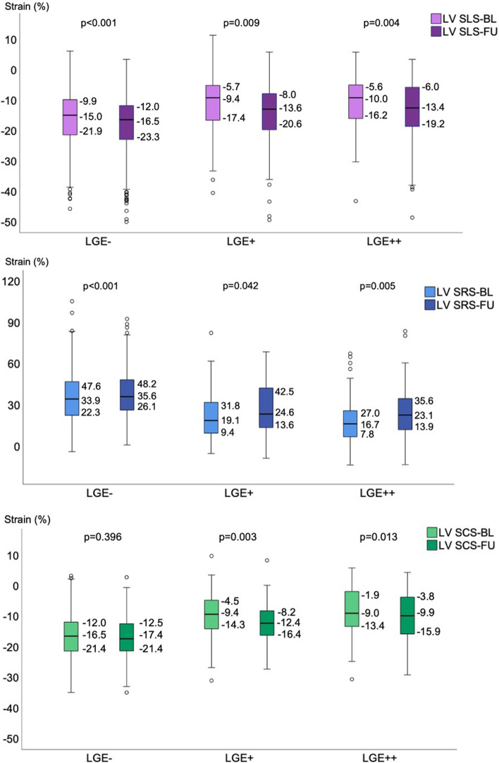
Strain is an important imaging parameter to determine myocardial deformation. This study sought to 1) assess changes in left ventricular strain and ejection fraction (LVEF) from acute to chronic ST-elevation myocardial infarction (STEMI) and 2) analyze strain as a predictor of late gadolinium enhancement (LGE). 32 patients with STEMI and 18 controls prospectively underwent cardiac magnetic resonance imaging. Patients were scanned 8 [Formula: see text] 5 days and six months after infarction (± 1.4 months). Feature tracking was performed and LVEF was calculated. LGE was determined visually and quantitatively on short-axis images and myocardial segments were grouped according to the LGE pattern (negative, non-transmural and transmural). Global strain was impaired in patients compared to controls, but improved within six months after STEMI (longitudinal strain from -14 ± 4 to -16 ± 4%, p < 0.001; radial strain from 38 ± 11 to 42 ± 13%, p = 0.006; circumferential strain from -15 ± 4 to -16 ± 4%, p = 0.023). Patients with microvascular obstruction showed especially attenuated strain results. Regional strain persisted impaired in LGE-positive segments. Circumferential strain could best distinguish between LGE-negative and -positive segments (AUC 0.73- 0.77). Strain improves within six months after STEMI, but remains impaired in LGE-positive segments. Strain may serve as an imaging biomarker to analyze myocardial viability. Especially circumferential strain could predict LGE.


Conflict of interest statement

The authors declare no competing interests.

Figures

Figure 1
Figure 1
Exemplary LGE-images (a), images of the strain analysis (b) and strain curves (c) of a patient with STEMI in the LAD territory. Abbreviations: STEMI ST-elevation myocardial infarction, LAD left anterior descending coronary artery, LV GLS left ventricular global longitudinal strain, LV GRS left ventricular global radial strain, LV GCS left ventricular global circumferential strain. Exemplary LGE-images (a) and strain curves (b) of a patient with STEMI in the LAD territory. In this exemplary study patient, all global strain values were impaired directly after myocardial infarction (LV GLS was −9%, LV GRS was 30% and LV GCS was −17%) and improved at follow-up. LGE showed a subendocardial pattern in the basal anterior wall, a transmural pattern midventricular and a semicircular pattern at the apex. Of the 16 myocardial segments, non-transmural LGE was noted in five and transmural LGE in three segments at baseline.
Figure 2
Figure 2
Box plots of the median (interquartile range) segmental strain results of patients at baseline and follow-up, grouped according to the extent of infarction. Abbreviations: LV SLS segmental longitudinal strain, LV SRS left ventricular segmental radial strain, LV SCS left ventricular segmental circumferential strain, LGE - myocardial segments without LGE, LGE + segments with non-transmural LGE, LGE +  + segments with transmural (> 50%) LGE. Box plots of the median (interquartile range) segmental strain results of patients at baseline and follow-up, grouped according to the extent of infarction. LV SLS, LV SRS and LV SCS improved from baseline to six-month follow-up. This improvement was observed in LGE − segments, as well as in LGE + und LGE +  + segments. Only LV SCS in LGE − segments did not change.
Figure 3
Figure 3
Results of the ROC-analysis on the diagnostic performance of segmental strain to predict LGE at baseline and follow-up. Abbreviations: LV GLS left ventricular global longitudinal strain, LV GRS left ventricular global radial strain, LV GCS left ventricular global circumferential strain, AUC area under the curve, s.e.: standard error. Results of the ROC-analysis on the diagnostic performance of segmental strain to predict LGE at baseline and follow-up. These receiver operating curves depict the ability of strain to predict LGE. The highest AUC was observed for LV SCS at baseline and follow-up. The ability of strain to predict LGE decreased by trend from baseline to follow-up.

References

    1. Roger VL. Epidemiology of heart failure. Circ. Res. 2013;113:646–659. doi: 10.1161/CIRCRESAHA.113.300268. - DOI - PMC - PubMed
    1. von Knobelsdorff-Brenkenhoff F, Pilz G, Schulz-Menger J. Representation of cardiovascular magnetic resonance in the AHA / ACC guidelines. J. Cardiovasc. Magn. Reson. 2017;19:70. doi: 10.1186/s12968-017-0385-z. - DOI - PMC - PubMed
    1. Konstam MA, Abboud FM. Ejection fraction: Misunderstood and overrated (Changing the Paradigm in Categorizing Heart Failure) Circulation. 2017;135:717–719. doi: 10.1161/CIRCULATIONAHA.116.025795. - DOI - PMC - PubMed
    1. Blessberger H, Binder T. Two dimensional speckle tracking echocardiography: clinical applications. Heart. 2010;96:2032–2040. doi: 10.1136/hrt.2010.199885. - DOI - PubMed
    1. Oyama-Manabe N, et al. Identification and further differentiation of subendocardial and transmural myocardial infarction by fast strain-encoded (SENC) magnetic resonance imaging at 3.0 Tesla. Eur. Radiol. 2011;21:2362–2368. doi: 10.1007/s00330-011-2177-4. - DOI - PubMed

Publication types