

Size variations in synaptic terminals among different types of photoreceptors and across the zebrafish retina
- PMID: 36587757
- PMCID: PMC9918681
- DOI: 10.1016/j.exer.2022.109377
Size variations in synaptic terminals among different types of photoreceptors and across the zebrafish retina
Abstract
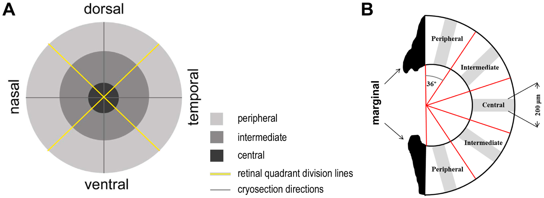
Photoreceptor synaptic terminals are responsible for transmitting visual information to downstream neurons. In vertebrate retinas, photoreceptor synaptic terminals are of different sizes and structures. The molecular mechanisms that underlie photoreceptor synaptic development are not clearly understood. Here, we have systematically examined the size variations in the synaptic terminals of cone and rod photoreceptors in the adult zebrafish retina. We reveal that the average cone pedicle sizes expand in the order of UV, blue, green, and red cones, echoing the increasing maximally sensitive wavelengths of the opsins expressed in the corresponding cone types. In addition, rod spherules are smaller than all cone pedicles. The terminals of each photoreceptor type also display distinct regional variations across the retina and between males and females. These findings establish the basis for using the zebrafish retina to study the molecular mechanisms that regulate the sizes and structures of photoreceptor terminals for proper visual functions.
Keywords: Pedicle; Photoreceptor; Ribbon synapse; Spherule; Synaptic terminal; Zebrafish.
Copyright © 2022 Elsevier Ltd. All rights reserved.
Figures


Similar articles
-
Spatiotemporal regulation of ATP and Ca2+ dynamics in vertebrate rod and cone ribbon synapses.Mol Vis. 2007 Jun 15;13:887-919. Mol Vis. 2007. PMID: 17653034 Free PMC article.
-
Synaptic plasticity and functionality at the cone terminal of the developing zebrafish retina.J Neurobiol. 2003 Sep 5;56(3):222-36. doi: 10.1002/neu.10243. J Neurobiol. 2003. PMID: 12884262
-
Ultrastructure of the distal retina of the adult zebrafish, Danio rerio.Tissue Cell. 2012 Aug;44(4):264-79. doi: 10.1016/j.tice.2012.04.004. Epub 2012 May 17. Tissue Cell. 2012. PMID: 22608306
-
The neuronal organization of the outer plexiform layer of the primate retina.Int Rev Cytol. 1984;86:285-320. doi: 10.1016/s0074-7696(08)60181-3. Int Rev Cytol. 1984. PMID: 6368448 Review.
-
The dynamic architecture of photoreceptor ribbon synapses: cytoskeletal, extracellular matrix, and intramembrane proteins.Vis Neurosci. 2011 Nov;28(6):453-71. doi: 10.1017/S0952523811000356. Vis Neurosci. 2011. PMID: 22192503 Free PMC article. Review.
References
-
- Braekevelt CR (1985). Photoreceptor fine structure in the archerfish. Am J Anat 173, 89–98. - PubMed
-
- Braekevelt CR (1994). Retinal photoreceptor fine structure in the great blue heron (Ardea herodias). Anat Histol Embryol 23, 281–292. - PubMed
-
- Braekevelt CR, Smith SA, and Smith BJ (1998). Photoreceptor fine structure in Oreochromis niloticus L. (Cichlidae; Teleostei) in light- and dark-adaptation. Anat Rec 252, 453–461. - PubMed
Publication types
MeSH terms
Grants and funding
LinkOut - more resources
Full Text Sources
