Review
. 2023 Jan 2;8(1):1.
doi: 10.1038/s41392-022-01259-6.

Targeting integrin pathways: mechanisms and advances in therapy

Affiliations
Review

Targeting integrin pathways: mechanisms and advances in therapy

Xiaocong Pang et al. Signal Transduct Target Ther. .

Abstract

Integrins are considered the main cell-adhesion transmembrane receptors that play multifaceted roles as extracellular matrix (ECM)-cytoskeletal linkers and transducers in biochemical and mechanical signals between cells and their environment in a wide range of states in health and diseases. Integrin functions are dependable on a delicate balance between active and inactive status via multiple mechanisms, including protein-protein interactions, conformational changes, and trafficking. Due to their exposure on the cell surface and sensitivity to the molecular blockade, integrins have been investigated as pharmacological targets for nearly 40 years, but given the complexity of integrins and sometimes opposite characteristics, targeting integrin therapeutics has been a challenge. To date, only seven drugs targeting integrins have been successfully marketed, including abciximab, eptifibatide, tirofiban, natalizumab, vedolizumab, lifitegrast, and carotegrast. Currently, there are approximately 90 kinds of integrin-based therapeutic drugs or imaging agents in clinical studies, including small molecules, antibodies, synthetic mimic peptides, antibody-drug conjugates (ADCs), chimeric antigen receptor (CAR) T-cell therapy, imaging agents, etc. A serious lesson from past integrin drug discovery and research efforts is that successes rely on both a deep understanding of integrin-regulatory mechanisms and unmet clinical needs. Herein, we provide a systematic and complete review of all integrin family members and integrin-mediated downstream signal transduction to highlight ongoing efforts to develop new therapies/diagnoses from bench to clinic. In addition, we further discuss the trend of drug development, how to improve the success rate of clinical trials targeting integrin therapies, and the key points for clinical research, basic research, and translational research.


Conflict of interest statement

The authors declare no competing interests.

Figures

Fig. 1
Fig. 1
Timeline of the historical milestone for the study of integrin receptors and their main antagonists and agents in the past four decades
Fig. 2
Fig. 2
The primary structure and representative conformations of integrins. a Organization of domains within the primary structures. b Arrangement of domains within the representative 3D crystal structure of integrins. c Conformational change of integrins: bent closed, extended–closed, and extended open conformations
Fig. 3
Fig. 3
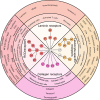
Classification, distribution, and ligands of integrins. The inner ring shows the 24 integrins that are composed of 17 α subunits and 8 β subunits. They are divided into four categories, namely, RGD-binding integrins, leukocyte cell-adhesion integrins, collagen-binding integrins, and laminin-binding integrins, according to their distribution, ligand specificity, and functions. The middle ring shows the distribution of integrins in different cell types. The outer ring indicates the ligands bound by different types of integrins
Fig. 4
Fig. 4
Schematic overview of integrin activation-associated signaling cascades. Integrin activation is regulated by multiple external signals, such as ECM, mechanotransduction or signaling from non-ECM ligands, including growth factor receptors, hormones, and small molecules, which is termed the “outside-in” mechanism. ECM or non-ECM ligand binding and force application results in integrin clustering and initiates downstream signaling to the actin cytoskeleton through recruited talin and vinculin, where actin can simultaneously pull on integrins and further in turn promote force generation. The “outside-in” mechanism then triggers various signaling cascades that ultimately result in cell survival, proliferation, cell spreading, and even tumorigenesis and metastasis. On the plasma membrane, there is also an “inside-out” mechanism, which regulates the displacement of intracellular integrin inhibitors and allows talin or kindlin binding to integrin β-tails, controlling integrin affinity for ECM components. For example, in neutrophils, both Talin-1 and Kindlin-3 are rapidly recruited to activate β2 integrins induced by extracellular chemokines binding to GPCR (G-protein-coupled receptor). Solid arrows indicate activation, the dotted line indicates recruiting, and the solid blunt end arcs indicate inhibitory effects
Fig. 5
Fig. 5
The expression and function of major integrins and their related cancer types and metastatic sites. The expression of integrins can vary considerably between normal and tumor tissue and is also associated with cancer types and organotrophic metastasis
Fig. 6
Fig. 6
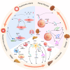
Roles of integrins in fibrosis processes in NASH, PH, and ADPKD. The lower part of the circle shows the role of integrins in liver fibrosis in NASH. In hepatic cells (HCs), activated integrin α9β1 is endocytosed by hepatocytes and secreted in the form of extracellular vesicles (EVs), which are further captured by MoMFs. Captured integrin α9β1 mediates MoMF adhesion to liver sinusoidal endothelial cells (LSECs) by binding to VCAM-1, which accelerates liver fibrosis. In HSCs, integrin α8β1 promotes liver fibrosis by activating TGF-β. The binding of integrin αvβ3 with OPN could promote laminin and α-SMA expression, which causes ECM accumulation and fibrosis progression. Integrin αvβ5 also binds with OPN and enhances liver fibrosis, but the underlying mechanism still needs to be clarified. In CD4 + T cells, the adhesion between integrin α4β7 and HC expressing MAdCAM-1 recruits CD4 + T cells to the liver, which induces liver inflammation and fibrosis. The left part of the circle shows the role of integrins in intimal fibrosis in PH. In the progression of PH, integrin α1, α8, αv, β1, and β3 are upregulated, and α5 is downregulated in PASMCs. Integrin α1 increases and α5 decreases the concentration of Ca2 + , promoting intimal fibrosis. The binding between integrin αvβ3 and OPN activates FAK signal transduction, which might be involved in the processes of vascular remodeling. The right part of the circle shows the role of integrins in renal fibrosis in ADPKD. Integrin αvβ3 expressed in renal tubular epithelial cells binds with periostin, activating TGF-β and promoting renal fibrosis. Binding between integrin αvβ3 and OPN is also involved in the renal fibrosis process, but the underlying mechanism is unclear. Renal tubular epithelial cells expressing integrin β1 enhance the expression of collagen, fibronectin, and α-SMA, which promote renal fibrosis
Fig. 7
Fig. 7
Main roles of integrins in the process of AS. Integrin signaling can affect multiple processes in AS, including endothelial dysfunction and activation, leukocyte homing to the plaque, smooth muscle cell migration, and thrombosis. In the process of endothelial cell activation, ox-LDL activates α5β1, induces the FAK/ERK/p90RSK pathway and promotes NF-κB signaling. Shear stress can activate αvβ3 and induce PAK activation by binding to fibronectin, thereby promoting NF-κB activation. Both ox-LDL and shear stress generated by blood flow mediate the increased expression of proinflammatory genes (ICAM-1 and VCAM-1) after integrin ligation. During the process of leukocyte homing to plaques, αxβ2 and α4β1 interact with VCAM-1 on the endothelial cell surface, and αxβ2 and αLβ2 interact with ICAM-1 to promote leukocyte adhesion. Integrins α4β1, α9β1 and αvβ3 on the surface of monocytes interact with osteopontin, which is expressed in atherosclerotic plaques, to promote monocyte migration and survival. Integrin αDβ2 is upregulated during macrophage foam cell formation. During vascular smooth muscle cell migration, αvβ3 binding with fibronectin, osteopontin, etc., mediates FAK activity and drives migration. In the process of thrombosis, integrins α2β1 and aIIbβ3 on platelets are involved in platelet adhesion, activation, aggregation, and thrombosis

References

    1. Hynes RO, Yamada KM. Fibronectins: multifunctional modular glycoproteins. J. Cell Biol. 1982;95:369–377. - PMC - PubMed
    1. Hynes RO. The emergence of integrins: a personal and historical perspective. Matrix Biol. 2004;23:333–340. - PMC - PubMed
    1. Kechagia JZ, Ivaska J, Roca-Cusachs P. Integrins as biomechanical sensors of the microenvironment. Nat. Rev. Mol. Cell Biol. 2019;20:457–473. - PubMed
    1. Moreno-Layseca P, Icha J, Hamidi H, Ivaska J. Integrin trafficking in cells and tissues. Nat. Cell Biol. 2019;21:122–132. - PMC - PubMed
    1. Zheng Y, Leftheris K. Insights into protein-ligand interactions in integrin complexes: advances in structure determinations. J. Med. Chem. 2020;63:5675–5696. - PubMed

Publication types