Review
. 2022 Dec 7;4(1):100360.
doi: 10.1016/j.xinn.2022.100360. eCollection 2023 Jan 30.

Myelin lipid metabolism and its role in myelination and myelin maintenance

Affiliations
Review

Myelin lipid metabolism and its role in myelination and myelin maintenance

Joseph A Barnes-Vélez et al. Innovation (Camb). .

Abstract

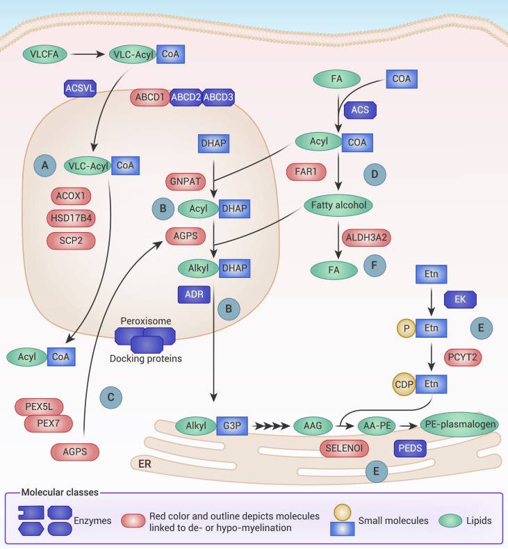
Myelin is a specialized cell membrane indispensable for rapid nerve conduction. The high abundance of membrane lipids is one of myelin's salient features that contribute to its unique role as an insulator that electrically isolates nerve fibers across their myelinated surface. The most abundant lipids in myelin include cholesterol, glycosphingolipids, and plasmalogens, each playing critical roles in myelin development as well as function. This review serves to summarize the role of lipid metabolism in myelination and myelin maintenance, as well as the molecular determinants of myelin lipid homeostasis, with an emphasis on findings from genetic models. In addition, the implications of myelin lipid dysmetabolism in human diseases are highlighted in the context of hereditary leukodystrophies and neuropathies as well as acquired disorders such as Alzheimer's disease.


Conflict of interest statement

The authors declare no competing interests.

Figures

Figure 1
Figure 1
Representative images of myelinated fibers and myelin sheath from mouse nerve (A) Left: transmission electron micrograph of myelinated and non-myelinated nerve fibers from cross-sectioned mouse sciatic nerve. Right: pseudo-colored overlay of the electron micrograph on the left. Red regions denote the myelin sheath, and yellow regions represent axon fibers. (B) High-magnification micrograph of mouse sciatic nerve to illustrate the multiple layers of myelin membrane that together compose the myelin sheath. (C) Pie chart delineating myelin lipid composition as molar percentage of total lipids as reported for bovine myelin from the CNS., Chol, cholesterol; Galc, galactocerebrosides; Sulf, sulfatides; Plas, plasmalogens; PC, phosphatidylcholine phospholipids; SM, sphingomyelin.
Figure 2
Figure 2
Cholesterol, sterol-response-element-binding protein (SREBP), and mammalian target of rapamycin (mTOR) pathways necessary in myelination (A) Farnesyl-diphosphate farnesyltransferase 1 (FDFT1) synthesizes squalene, a key intermediate in cholesterol synthesis. (B) Low-density lipoprotein receptor-related protein 1 (LRP1) enables endocytosis of lipoprotein particles, which deliver exogenous cholesterol. (C) Sterol cleavage-activating protein (SCAP), in response to decreased ER cholesterol, shuttles the SREBPs to the Golgi apparatus for cleavage-mediated activation by site-1 and site-2 proteases (S1P and S2P). (D) Elevated ER cholesterol induces SCAP to associate with and be inhibited by insulin-induced gene protein (Insig), reducing SCAP-mediated SREBP2 activation. (E) Quaking (QKI) is a critical co-activator for SREBP2 in OLs that is necessary for transcription of cholesterol-synthesizing genes. (F) mTOR, regulatory-associated protein of mTOR (Raptor), and mTOR-associated protein, LST8 homolog (mLst8) are subunits of the mTORC1 complex that increase SREBP activity through multiple mechanisms. One reported mechanism includes inhibiting the flux of autophagolysosome-derived cholesterol to the ER and preventing cholesterol-mediated inhibition on SREBP activation. OL, oligodendrocyte.
Figure 3
Figure 3
Fatty acid, galactocerebroside, and sulfatide synthesis pathways implicated in myelination and myelin maintenance (A) Condensation of serine and palmitoyl-CoA by serine palmitoyltransferase (SPT) initiates Cer synthesis, which is then completed through the activity of ceramide synthase (Cers) and dihydroceramide desaturase (DEGS). (B) Ceramide galactosyl-transferase (CGT)-mediated addition of galactose to Cer generates GalC. Following translocation to the Golgi, addition of sulfate to GalC by cerebroside sulfotransferase (CST) forms Sulf. Conversely, Cer taken to the Golgi can be glycosylated to GlcC by UDP-glucose ceramide glucosyltransferase (UGCG), which can be further glycosylated into complex glycosphingolipids including globosides and gangliosides. (C) Fatty acid 2-hydroxylase (FA2H) catalyzes FA hydroxylation. Hydroxylated FA (2OH-FA) are used as substrates by sphingolipid-synthesizing enzymes to generate hydroxylated sphingolipids. (D) The rich heterogeneity of sphingolipids stems from the multiple species of FA used as substrates for dihydrosphingosine acylation, including saturated (SFA), monounsaturated (MUFA), and polyunsaturated (PUFA) FA. Elongation of endogenous or diet-derived FA requires fatty acid elongases (ELOVL) and hydroxyacyl dehydratases (HACD). FA desaturation depends on FA desaturases, including FADS and SCD. (E) Fatty acid synthase (FASN) generates palmitate, a principal source for endogenous SFA and MUFA. (F) The nuclear receptors peroxisome proliferator-activated receptor β (PPARβ) and retinoic X receptor α (RXRα), following co-activation by quaking (QKI), drive transcription of the FA biosynthesis pathway. FA, fatty acid; Cer, ceramide; GalC, galactocerebroside; Sulf, sulfatides; GlcC, glucocerebroside.
Figure 4
Figure 4
Elements of peroxisome, plasmalogen, and phospholipid metabolism required for myelin integrity (A) Oxidation of very-long-chain fatty acids (VLCFAs) occurs within peroxisomes. VLC-acyl-CoA are imported by ATP-binding cassette transporter subfamily D (ABCD) transporters, principally ABCD1. VLC-acyl-CoA are then oxidized into shorter-chain acyl-CoA by a series of reactions requiring acyl-CoA oxidase (ACOX1), 17-β-hydroxysteroid dehydrogenase IV (HSD17B4), and sterol carrier protein 2 (SCP2). (B) Initial steps for plasmalogen synthesis require peroxisomal enzymes. Glycerone phosphate-O-acyltransferase (GNPAT) synthesizes 1-acyl-DHAP, which is converted to 1-alkyl-O-DHAP by alkylglycerone phosphate synthase (AGPS). 1-Alkyl-O-DHAP is reduced by alkyl DHAP reductase (ADR) to alkyl-G3P, which passes to the ER to complete plasmalogen synthesis. (C) Peroxisome matrix proteins, including AGPS, require carrier proteins, such as PEX7 and the PEX5L isoform, to enter peroxisomes. (D) Fatty alcohols derived from fatty acyl-CoA reductase 1 (FAR1) are utilized for plasmalogen synthesis. (E) Most plasmalogens in myelin exist as phosphatidylethanolamine lipids (PE-plasmalogens). Ethanolamine (Etn) is phosphorylated by ethanolamine kinase (EK) and coupled with CDP by ethanolamine phosphate cytidylyltransferase (PCYT2). Selenoprotein I (SELENOI) converts 1-O-alkyl-2-acylglycerol (AAG) into 1-O-alkyl-2-acyl-GPE (AA-PE) using PCYT2-derived CDP-Etn. AA-PE is then converted to PE-plasmalogen by plasmanylethanolamine desaturase (PEDS). (F) Balance of fatty acids and fatty alcohols is maintained in part by members of the aldehyde dehydrogenase (ALDH) family, including ALDH3A2. CDP, cytidine diphosphate; DHAP, dihydroxyacetone phosphate; ACSVL, very-long-chain acyl-CoA-synthase; ACS, acyl-CoA synthase.
Figure 5
Figure 5
Myelin disruption in leukodystrophies and Alzheimer’s disease (A) Leukodystrophy-associated lipid dysregulation occurs from mutations in genes that contribute to lipid synthesis or catabolism (see Table 1). (B) With aging, there is a decline with brain lipids enriched in myelin and a decrease in myelin renewal. (C) APP/PS1 mice experience demyelination, which exacerbates AD-like memory deficits. Clemastine treatment or conditional knockout (cKO) of M1R in OPCs restored myelin renewal and ameliorated cognitive decline.

Similar articles

Cited by

References

    1. Tasaki I. The electro-saltatory transmission of the nerve impulse and the effect of narcosis upon the nerve fiber. American Journal of Physiology-Legacy Content. 1939;127:211–227.
    1. Huxley A.F., Stämpfli R. Evidence for saltatory conduction in peripheral myelinated nerve fibres. J. Physiol. 1949;108:315–339. - PubMed
    1. Pease-Raissi S.E., Chan J.R. Building a (w)rapport between neurons and oligodendroglia: reciprocal interactions underlying adaptive myelination. Neuron. 2021;109:1258–1273. - PMC - PubMed
    1. Xin W., Chan J.R. Myelin plasticity: sculpting circuits in learning and memory. Nat. Rev. Neurosci. 2020;21:682–694. - PMC - PubMed
    1. Yeung M.S.Y., Zdunek S., Frisén J. Dynamics of oligodendrocyte generation and myelination in the human brain. Cell. 2014;159:766–774. - PubMed

LinkOut - more resources