Unraveling rare form of adult-onset NIID by characteristic brain MRI features: A single-center retrospective review
- PMID: 36588885
- PMCID: PMC9798416
- DOI: 10.3389/fneur.2022.1085283
Unraveling rare form of adult-onset NIID by characteristic brain MRI features: A single-center retrospective review
Abstract
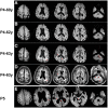
Adult-onset neuronal intranuclear inclusion disease (NIID) is a rare neurodegenerative disorder with high clinical heterogeneity. Previous studies indicated that the high-intensity signals in the corticomedullary junction on diffusion-weighted imaging (DWI) on brain MRI, known as the "ribbon sign," could serve as a strong diagnostic clue. Here we used the explorative approach to study the undiagnosed rate of adult-onset NIID in a single center in China via searching for the ribbon sign in picture archive and communication system (PACS) and report the clinical and radiological features of initially undiagnosed NIID patients. Consecutive brain MRI of 21,563 adult individuals (≥18 years) in the PACS database in 2019 from a tertiary hospital were reviewed. Of them, 4,130 were screened out using the keywords "leukoencephalopathy" and "white matter demyelination." Next, all 4,130 images were read by four neurologists. The images with the suspected ribbon sign were reanalyzed by two neuroradiologists. Those with the ribbon sign but without previously diagnosed NIID were invited for skin biopsy and/or genetic testing for diagnostic confirmation. The clinical features of all NIID patients were retrospectively reviewed. Five patients with high-intensity in the corticomedullary junction on DWI were enrolled. Three patients were previously diagnosed with NIID confirmed by genetic or pathological findings and presented with episodic encephalopathy or cognitive impairment. The other two patients were initially diagnosed with limb-girdle muscular dystrophy (LGMD) with rimmed vacuoles (RVs) and normal pressure hydrocephalus (NPH) in one each. Genetic analysis demonstrated GGC repeat expansion in the NOTCH2NLC gene of both, and skin biopsy of the first patient showed the presence of intranuclear hyaline inclusion bodies. Thus, five of the 21,563 adult patients (≥18 years) were diagnosed with NIID. The distinctive subcortical high-intensity signal on DWI was distributed extensively throughout the lobes, corpus callosum, basal ganglia, and brainstem. In addition, T2-weighted imaging revealed white matter hyperintensity of Fazekas grade 2 or 3, atrophy, and ventricular dilation. Distinctive DWI hyperintensity in the junction between the gray and white matter can help identify atypical NIID cases. Our findings highly suggest that neurologists and radiologists should recognize the characteristic neuroimaging pattern of NIID.
Keywords: MRI; leukoencephalopathy; myopathy; neuronal intranuclear inclusion disease (NIID); ribbon sign.
Copyright © 2022 Li, Wang, Zhu, Xiao, Gu, Yu, Deng, Sun and Wang.
Conflict of interest statement
The authors declare that the research was conducted in the absence of any commercial or financial relationships that could be construed as a potential conflict of interest.
Figures


Similar articles
-
Identifying patients with neuronal intranuclear inclusion disease in Singapore using characteristic diffusion-weighted MR images.Neuroradiology. 2019 Nov;61(11):1281-1290. doi: 10.1007/s00234-019-02257-2. Epub 2019 Jul 11. Neuroradiology. 2019. PMID: 31292692
-
Neuronal intranuclear inclusion disease in patients with adult-onset non-vascular leukoencephalopathy.Brain. 2022 Sep 14;145(9):3010-3021. doi: 10.1093/brain/awac135. Brain. 2022. PMID: 35411397
-
NOTCH2NLC GGC Repeat Expansion in Patients With Vascular Leukoencephalopathy.Stroke. 2023 May;54(5):1236-1245. doi: 10.1161/STROKEAHA.122.041848. Epub 2023 Mar 21. Stroke. 2023. PMID: 36942588
-
A case report of neuronal intranuclear inclusion disease and literature review.BMC Neurol. 2024 Dec 20;24(1):488. doi: 10.1186/s12883-024-03997-2. BMC Neurol. 2024. PMID: 39707256 Free PMC article. Review.
-
Neuronal intranuclear inclusion disease: two case report and literature review.Neurol Sci. 2021 Jan;42(1):293-296. doi: 10.1007/s10072-020-04613-0. Epub 2020 Aug 25. Neurol Sci. 2021. PMID: 32839883 Review.
Cited by
-
Not your usual neurodegenerative disease: a case report of neuronal intranuclear inclusion disease with unconventional imaging patterns.Front Neurosci. 2023 Aug 10;17:1247403. doi: 10.3389/fnins.2023.1247403. eCollection 2023. Front Neurosci. 2023. PMID: 37638306 Free PMC article.
-
Neuronal Intranuclear Inclusion Disease with NOTCH2NLC GGC Repeat Expansion: A Systematic Review and Challenges of Phenotypic Characterization.Aging Dis. 2024 Feb 16;16(1):578-97. doi: 10.14336/AD.2024.0131-1. Online ahead of print. Aging Dis. 2024. PMID: 38377026 Free PMC article.
-
A case of neuronal intranuclear inclusion disease (NIID) presenting with hydrocephalus-like clinical features: case report.BMC Neurol. 2025 Feb 6;25(1):51. doi: 10.1186/s12883-025-04056-0. BMC Neurol. 2025. PMID: 39915717 Free PMC article.
References
LinkOut - more resources
Full Text Sources

