

Clinical characteristics and management experience of schwannoma in extremities: Lessons learned from a 10-year retrospective study
- PMID: 36588891
- PMCID: PMC9797853
- DOI: 10.3389/fneur.2022.1083896
Clinical characteristics and management experience of schwannoma in extremities: Lessons learned from a 10-year retrospective study
Abstract
Introduction: Schwannomas are the most common neoplastic lesions of the peripheral nerves when growing on the extremities, they usually have adverse effects on patients due to the exposed and functional nature of the region.
Methods: In the present single-center retrospective study, we included all patients with pathologically confirmed schwannoma located in extremities between 2011 and 2021 totaling 183 patients. Data on gender, age, duration history, clinical presentation, occurrence region, nerve affiliation, imaging data, modus operation, mass volume, immunohistochemistry, postoperative neurological function, and recurrence were collected.
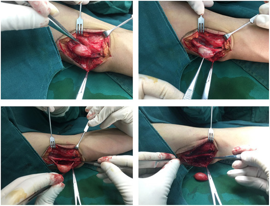
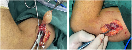
Results: As in previous studies, patients were predominantly middle-aged with a mean age of 49.5, without gender preference and a male-to-female ratio of 1.2:1. Most patients are first seen for this disease, and only five of them are recurrent. The majority presented with an isolated (91.26%), asymptomatic (37.7%) mass, with tenderness (34.97%) being the second frequent complaint. 60% of lesions occurred in the upper extremity, more commonly on the left side (55.26%) than the right. The average duration of onset was 47.50 months. MRI is more sensitive for neurogenic tumors than ultrasound, as it owns 78.93% correct. In immunohistochemistry, the top three markers for positive labeling schwannoma are S-100 (98.95%), Ki67 (98.68%) and β-Catenin. 98.36% of patients underwent complete resection of the lesion, of which 14.44% required partial sacrifice of the nerve fibers. Thanks to the application of intraoperative peripheral nerve microscopic operation, only 6 patients showed symptoms of postoperative nerve injury, and 3 of them received second surgery. Intraoperative microscopic manipulation, preservation of the main nerve, and the need for reconstruction of the affected nerve fibers are some of the points worth noting.
Discussion: In summary, the possibility of schwannoma should not be overlooked in the identification of masses that occur in the upper extremities of the middle-aged population. Preoperative ultrasound and MR are useful for determining the nature of the mass, and S100, Ki67, and β-Catenin are sensitive to it. Surgical resection can achieve satisfying functional results and a low risk of nerve injury.
Keywords: diagnosis; extremity; microsurgery; prognosis; retrospective study; schwannoma.
Copyright © 2022 Zhou, Yao, Dong, Alhaskawi, Wang, Lai, Ezzi, Kota, Abdulla and Lu.
Conflict of interest statement
The authors declare that the research was conducted in the absence of any commercial or financial relationships that could be construed as a potential conflict of interest.
Figures


References
-
- Institute NC . Schwannoma Was Originally Published by the National Cancer Institute 2020 (accessed July 30, 2020).
LinkOut - more resources
Full Text Sources
Research Materials