Review
. 2022 Dec 9:30:e00184.
doi: 10.1016/j.fawpar.2022.e00184. eCollection 2023 Mar.

Endemic Paragonimus kellicotti infections in animals and humans in USA and Canada: Review and personal perspective

Affiliations
Review

Endemic Paragonimus kellicotti infections in animals and humans in USA and Canada: Review and personal perspective

J P Dubey. Food Waterborne Parasitol. .

Abstract

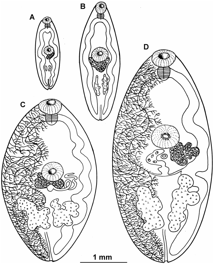
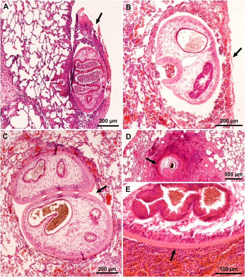
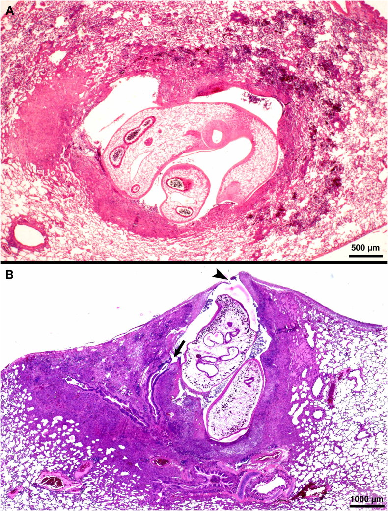
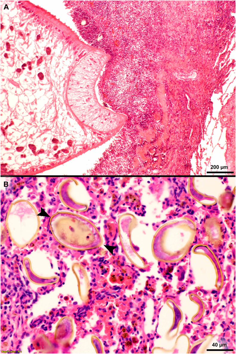
Infections with the lung fluke, Paragonimus kellicotti, have been diagnosed in a variety of domestic and wild animals and humans in USA and Canada. Although there are many species of Paragonimus in other parts of the world; P. kellicotti is the only species definitively diagnosed in USA and Canada. Fresh water snails (several species) and crayfish (mainly Orconectes spp.) are its intermediate hosts. Humans and animals become infected with P. kellicotti only by ingesting metacercariae encysted in the heart of crayfish. After ingestion, the fluke penetrates intestinal wall, enters peritoneal cavity, and reaches pleural cavity by direct penetration of diaphragm, 2-3 weeks post inoculation (p.i.). Young flukes penetrate lungs and become encysted in pulmonary tissue, often in pairs. Time to maturity is around 4-7 weeks p.i. Eggs are coughed up, swallowed, and are excreted in feces. Although the parasite has been known for more than a century, there has been an upsurge of human infections in the USA. Here, I review P. kellicotti infections in naturally infected hosts. Pathogenesis, diagnosis, and treatment in parasite-free cats and dogs experimentally infected P. kellicotti are reviewed to shed light on the pathogenesis of human paragonimiasis. Problems and challenges facing diagnosis of paragonimiasis, especially non-pulmonary infections, are discussed. Fluke stages are deposited in Smithsonian Museum.

Keywords: Animals; Diagnosis; Epidemiology; Humans; Life cycle; Paragonimus kellicotti; Treatment.


Conflict of interest statement

The authors declare that they have no known competing financial interests or personal relationships that could have appeared to influence the work reported in this paper.

Figures

Unlabelled Image
Graphical abstract
Fig. 1
Fig. 1
Life cycle of Paragonimus kellicotti in cats. (From Dubey et al., 1978a).
Fig. 2
Fig. 2
Environmental stages of Paragonimus kellicotti. (A, B) Eggs in feline feces, using sedimentation technique (A) and salt flotation technique (B). Unstained. Eggs are multicelled, operculated (arrowheads), and golden brown. There is a thickening (arrow) at the pole opposite the operculum. Paragonimus eggs become distorted in hypertonic salt solution, leaving only the eggshell. (C) Crayfish (Orconectes sp.), the second intermediate host of P. kellicotti, dissected to show the white gelatinous heart. Metacercaria (circled) is embedded in the heart of crayfish. (D) Metacercaria removed from the crayfish. Note dark excretory system (e) and the white ceca (c). (E) Young fluke squeezed out of metacercaria. Note oral (os), ventral sucker (vs, partly obscured), excretory vesicle (e), and 2 ceca (c). (Figs. A, B, C from Dubey et al., 1978a; Figs. D and E, courtesy of Dr. Peter Fisher, University of Washington, St. louis, Missouri, USA). (For interpretation of the references to colour in this figure legend, the reader is referred to the web version of this article.)
Fig. 3
Fig. 3
Paragonimus kellicotti stages recovered from experimentally infected cats. Flukes were fixed under moderate pressure and stained with Semichon's carmine as described (Stromberg and Dubey, 1978). (A) Day 7 p.i. (B) Day 14 p.i. (C) Day 21 p.i. (D) Day 29 p.i. (E) Day 34 p.i. Flukes at days 7 and 14 p.i.appear morphologically similar.
Fig. 4
Fig. 4
Line drawings of Paragonimus kellicotti from experimentally infected cats. The drawings were made from flukes shown in Fig. 3. Fluke at day 7 p.i. is like the fluke released from the metacercaria (not shown). (A) Fluke at day 14 p,i., female genital appeared just below ventral sucker (also called acetabulum) and testicular primordia were seen towards the excretory pore. (B) Fluke at day 21 p,i, main vitelline ducts have formed. (C) Fluke at day 29 p.i., the tubular uterus, and shell glands formed. (D) Fluke sat day 34 p.i., a few eggs had formed. (From Stromberg and Dubey, 1978).
Fig. 5
Fig. 5
Pathogenesis of pulmonary paragonimiasis in experimentally infected cats. Histological sections of lungs. HE-stain. (A) Penetration of pleura by immature fluke. Note inflammatory exudate (arrow) with fibrin, eosinophils, extravasated erythrocytes covering the fluke. Day 10 p.i. (B) Recently penetrated fluke in subpleural cavity of pulmonary parenchyma. The pleural breakage (arrow) is healing. The fluke is feasting on host tissue. Day 14 p.i. (C) Paired immature flukes within lung parenchymal cavity (arrow). (D, E) Immature fluke with intense eosinophilic inflammation and necrotic tissue (arrows) surround the fluke. Day 21 p.i. (From Hoover and Dubey, 1978).
Fig. 6
Fig. 6
Cystic lesions in lungs of cats fed Paragonimus kellicotti metacercariae. Histological sections of lungs. HE-stain. (A) Early stage of cyst formation. Two immature flukes are surrounded by hemorrhagic exudate containing eosinophils and other inflammatory contents. Day 29 after feeding 25 metacercariae. (B) A cyst with a very thin or broken pleural surface (arrowhead) and a communication with bronchus (arrow). Two mature flukes are surrounded by fibrous tissue, exudate, and granulomatous tissue. Eggs are present in surrounding tissue but not visible at this magnification. Day 39 after feeding 34 metcercariae.
Fig. 7
Fig. 7
Microscopic appearance of Paragonimus kellicotti in histological sections of lungs. HE-stain, day 70 after feeding 25 metacercariae. (A) Cat. Anterior end of a fluke attached to granulomatous tissue. The cuticular spines of the fluke probably irritate the host tissue provoking granulomatous response. (B) Dog. Several golden-brown eggs are trapped in alveolar tissue. One of the eggs is cut longitudinally (opposing arrowheads) with operculum at one end and a slight thickening at the narrow end. (For interpretation of the references to colour in this figure legend, the reader is referred to the web version of this article.)
Fig. 8
Fig. 8
Gross appearance of lesions in lungs of cats fed Paragonimus kellicotti metacercariae. Unstained. (A) Early hemorrhagic lesions in a cat day 23 after feeding 50 metacercariae. More lesions are present in caudal lobes (near penetration site in diaphragm) than in other lobes. The number of hemorrhagic spots (arrowhead) exceed the number of metacercaria fed indicating repeated penetration of flukes searching for a site to settle. Note one pleural plaque (arrow), probable the site where fluke has successfully penetrated. (B) Dorsal view of early hemorrhagic appearance of cysts (arrow) in a cat day 29 after feeding 50 metacercariae. Arrowheads point to early penetration sites. (C) Ventral view of lungs of a cat 56 days after feeding 26 metacercariae. Bronchial lymph node (arrowhead) is enlarged, the mediastinum is colored brownish (shaded area) due to eggs, and cysts are also greyish (arrow). (D) Incised cyst revealing two flukes (arrows) in a cat day 263 after feeding 15 metacercaria (From Dubey et al., 1978a).
Fig. 9
Fig. 9
Lungs of a P. kellicotti infected cat, 21 days after treating with albendazole (50 mg/kg body weight). Treatment of cat was started on day 101 after feeding 25 metacercariae and therapy was continued for 21 days. HE-stain. (A) Note a dead fluke (arrow) and a hemorrhagic tract (opposing arrowheads) indicating probably the tract left by a fluke that has existed from the lung. (B) A fluke (arrow) exiting from pleura.

References

    1. Ah H.S., Chapman W.L., Jr. Extrapulmonary granulomatous lesions in canine paragonimiasis. Vet. Parasitol. 1976;2:251–258.
    1. Alden C.L., Gay S., Adkins A. Pulmonary trematodiasis in a cat: a case report. Vet. Med. Small Anim. Clin. 1980;75:612–617. - PubMed
    1. Ameel D.J. More data on the lung fluke, Paragonimus, in North America. Science. 1931;74:493–494. - PubMed
    1. Ameel D.J. The muskrat, a new host for Paragonimus. Science. 1932;75:382. - PubMed
    1. Ameel D.J. Life history of the North American lung fluke of mammals. J. Parasitol. 1932;18:264–268.

LinkOut - more resources