. 2021 Nov 1;13(4):331-346.
doi: 10.1007/s12609-021-00435-x.

Uncommon Tumors and Uncommon Presentations of Cancer in the Breast

Affiliations

Uncommon Tumors and Uncommon Presentations of Cancer in the Breast

Marina J Corines et al. Curr Breast Cancer Rep. .

Abstract

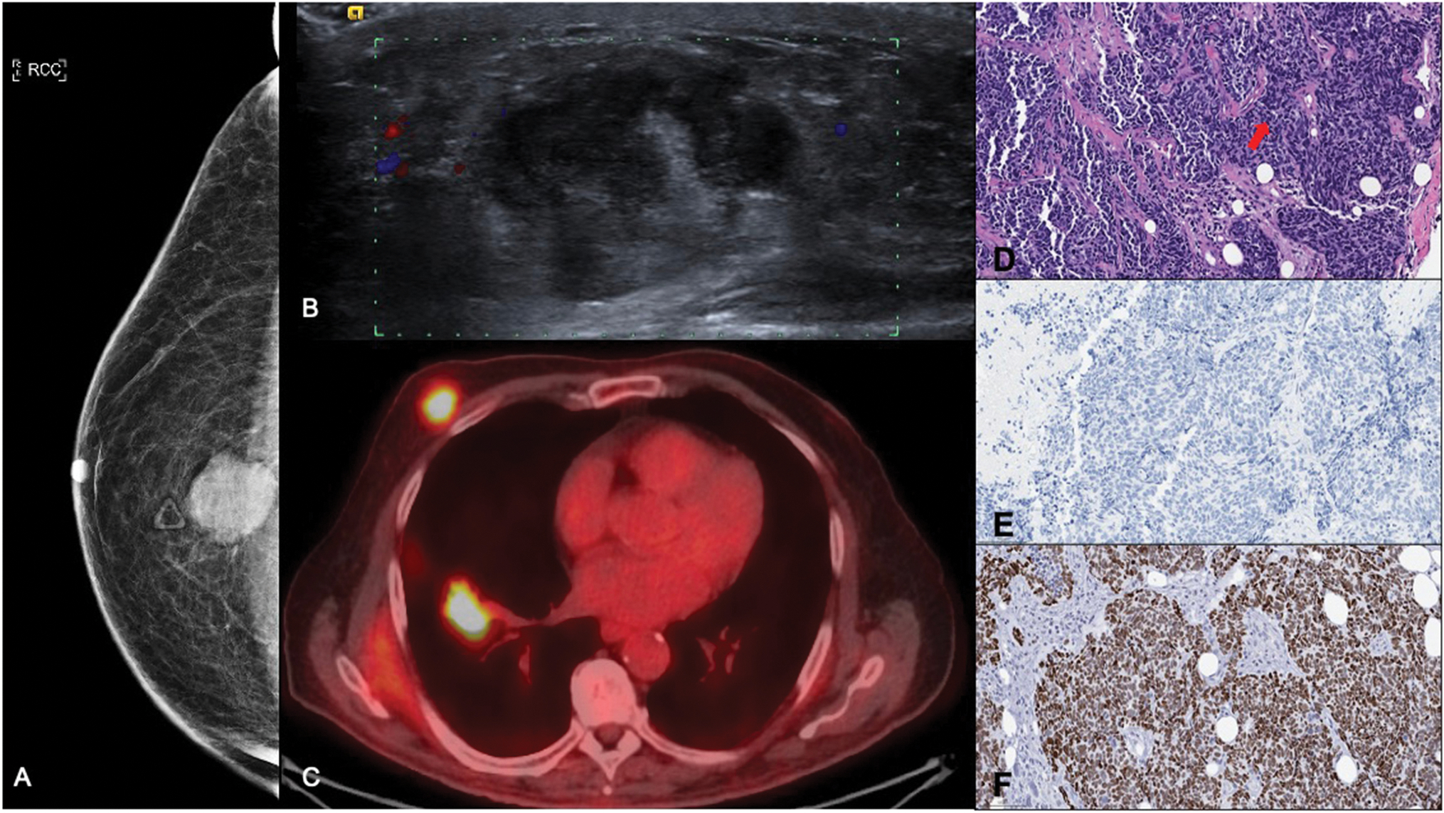
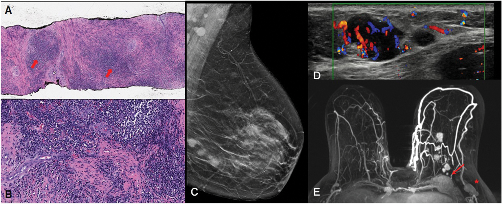
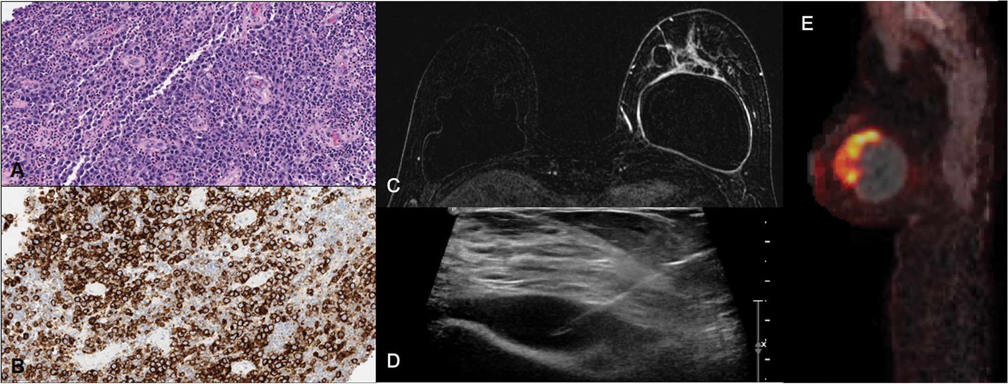
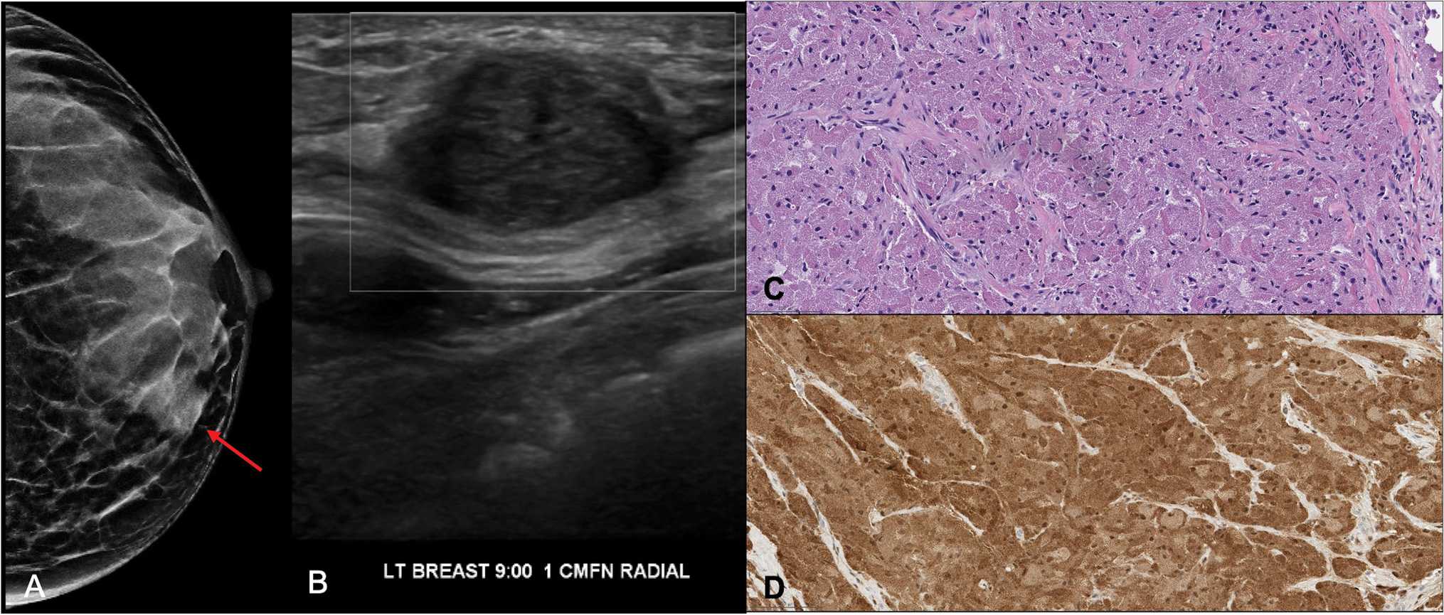
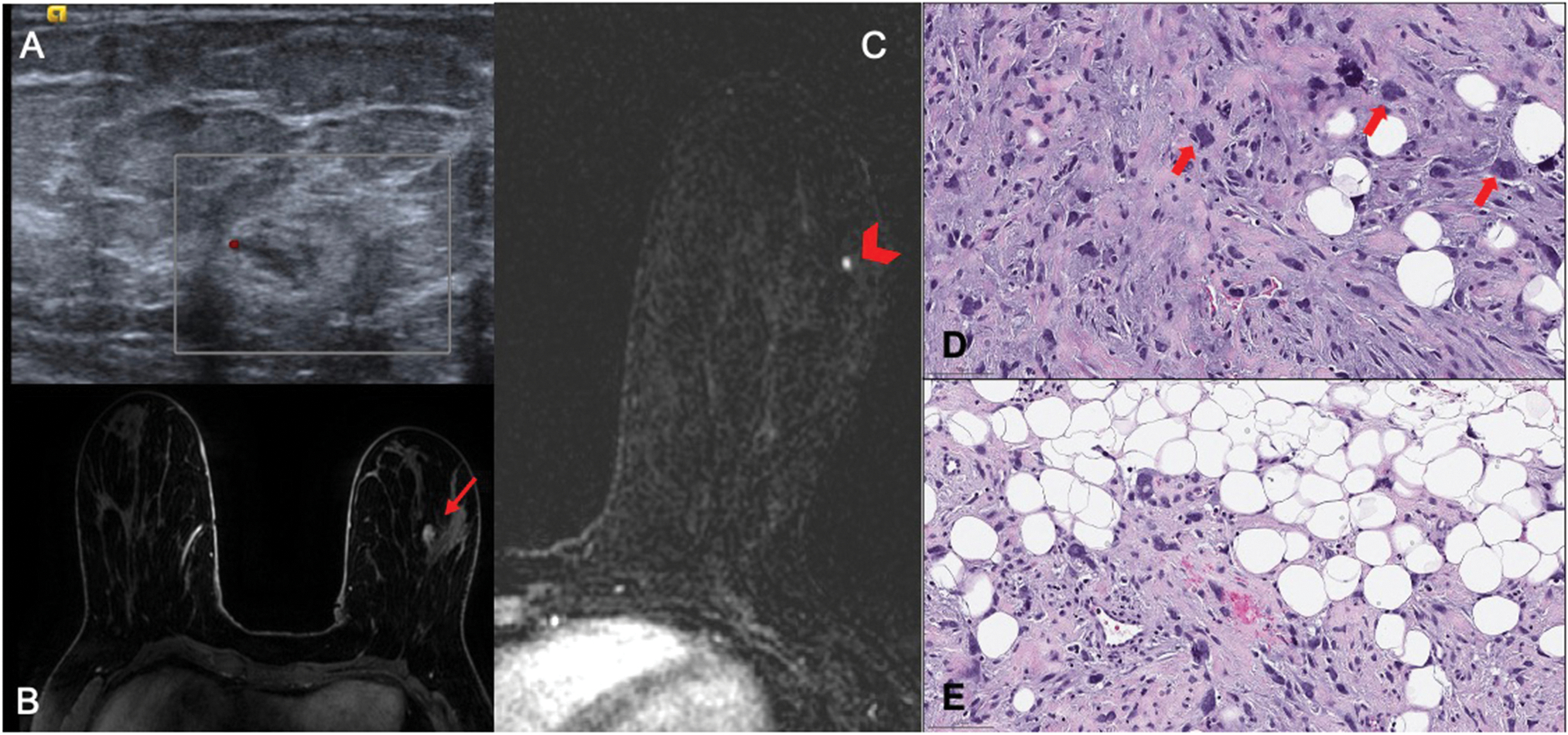
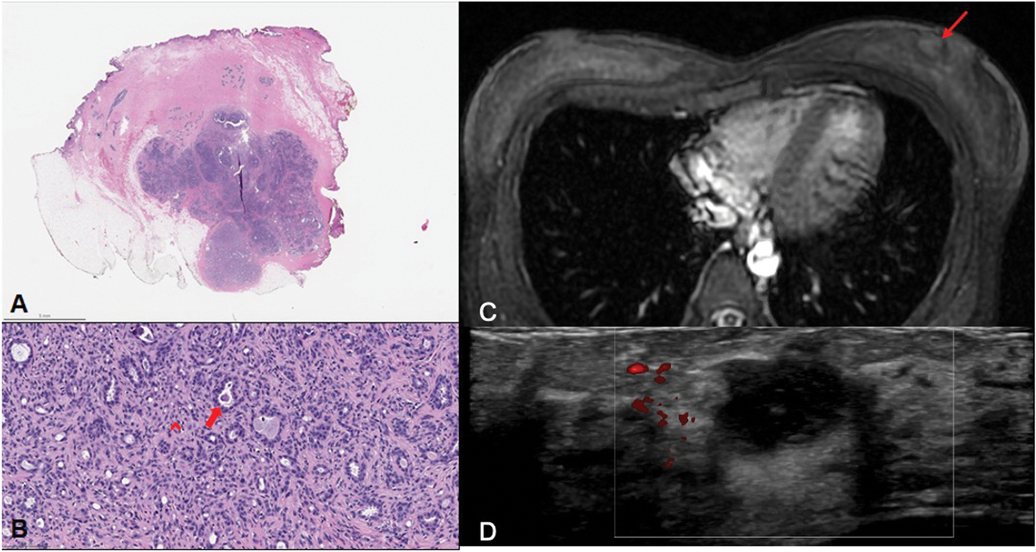
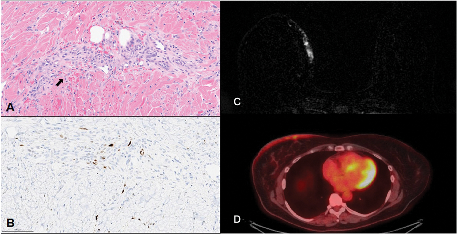
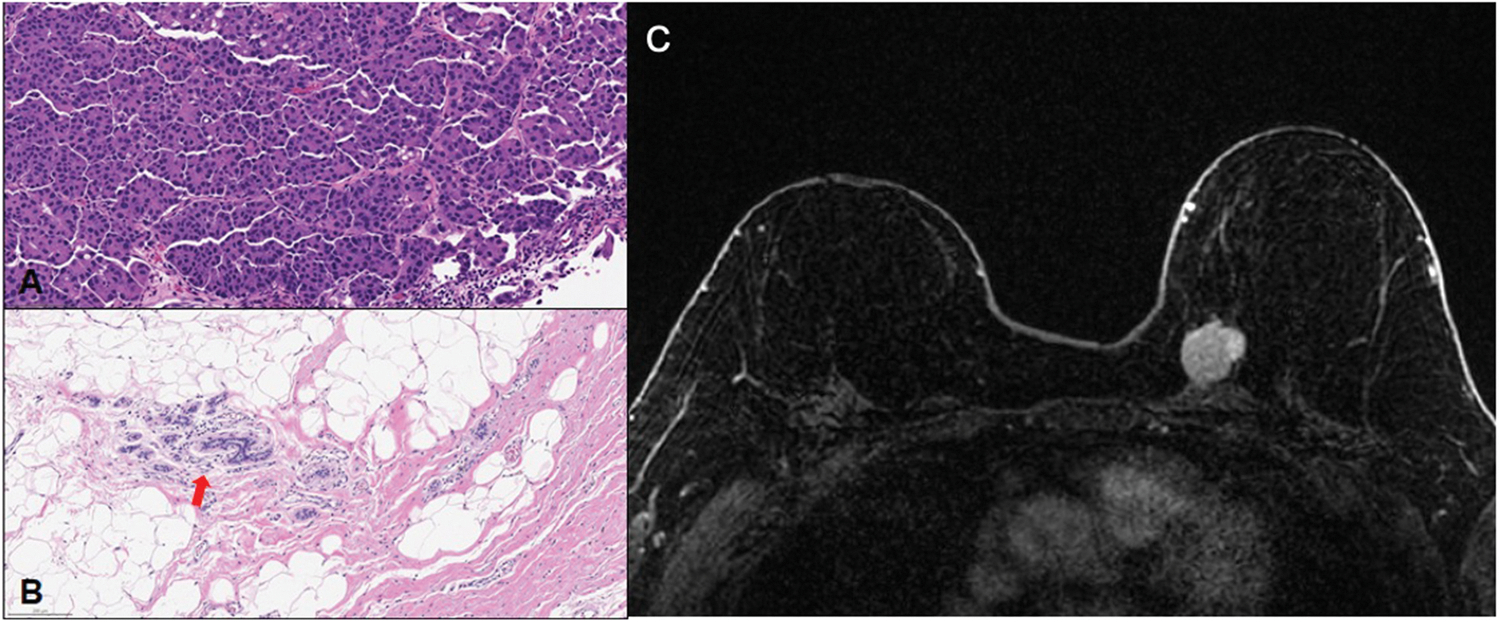
Purpose of review: The purpose is to present a case series of rare diagnoses and unusual presentations of breast lesions with radiologic-pathologic correlation from a major cancer center, and to review the recent literature on each entity with a focus on radiology-pathology concordance. We present our findings and experience from cases of metastatic small cell lung carcinoma to the breast, IgG-4 related breast disease, breast implant associated anaplastic large cell lymphoma, granular cell tumor, pleomorphic sarcoma, adenomyoepithelioma, post-radiation angiosarcoma, and breast carcinoma after risk-reducing total mastectomy.

Recent findings: It is essential for physicians to have knowledge of rare breast diagnoses and unusual breast disease presentations to formulate a complete differential diagnosis, recognize radiological-pathological concordance of these entities and provide appropriate patient care.

Summary: Current literature on these rare described entities exists mainly as case reports, case series and small-scale studies. By sharing our findings, we hope to educate trainees in radiology, pathology and other fields across the continuum of care in radiologic-pathologic correlation, while also augmenting the existing literature on these rare entities.

Keywords: Extramammary metastasis to breast; IgG4 related disease; adenomyoepithelioma; breast implant associated anaplastic large cell lymphoma; granular cell tumor; risk-reducing total mastectomy; sarcoma.


Conflict of interest statement

Conflict of Interest Marina Corines, Melissa Krystel-Whittemore, and Melissa Murray declare that they have no conflict of interest. Victoria Mango reports personal fees from Bayer Healthcare, personal fees from Koios Medical, outside the submitted work.

Figures

Figure 1:
Figure 1:. Metastatic small cell lung cancer to the breast initially diagnosed as poorly differentiated triple negative male breast cancer.
A) Diagnostic mammography RCC view demonstrates a right breast 5:00 axis posterior depth round, indistinct, hyperdense mass measuring 3.2 cm in the region of palpable concern delineated by triangular marker. B) Targeted ultrasound demonstrates a corresponding irregular hypoechoic mass with peripheral vascularity, abutment of the pectoralis muscle, and surrounding edema, which correlated with the palpable abnormality. C)18F-FDG PET/CT for initial breast cancer staging fused axial image demonstrates hypermetabolic right breast 5:00 axis mass (SUVmax 9.8), as well as a hypermetabolic right lower lobe pulmonary mass (SUVmax 13.9) measuring 3.1 cm, suspicious for malignancy. D) Hematoxylin and eosin (H&E, 200x) stained slide from percutaneous breast biopsy shows a poorly differentiated carcinoma with spindle cell features (arrow). While this may represent breast carcinoma, the absence of in situ component and ER/PR/HER2 negativity suggest further work up is warranted. E) GATA3 immunohistochemical staining of the tumor was negative, which is often positive in breast carcinoma. F) TTF1 immunohistochemical staining shows strong positivity of the tumor cells, supporting the diagnosis of metastatic small cell carcinoma.
Figure 2.
Figure 2.. IgG4-related disease of the breast.
A) Low-power (40x) of the core from left breast percutaneous biopsy (H&E stain) shows dense lymphoplasmacytic inflammation (arrows) in a background of haphazard fibrosis. B) High power (200x) image shows dense lymphoplasmacytic inflammation with numerous eosinophils. C) Mammography LMLO view demonstrates multiple left lower breast dense circumscribed masses in a ductal distribution spanning 4.7 cm and prominent left axillary lymph nodes. D) Ultrasound with color flow demonstrates multiple left breast masses in the 7:00 axis 8–10 cm from the nipple measuring up to 2.9 × 2.7 cm with associated vascularity. Abnormal appearing left axillary lymph nodes were also sonographically noted (not shown). E) Breast MRI T1-fat suppressed post contrast subtraction maximum intensity projection (MIP) image demonstrates multiple heterogeneously enhancing masses in the left breast involving the chest wall (arrow) and multiple ribs, corresponding to the sonographic masses, and left axillary adenopathy (*). Subsequent laboratory results demonstrated an elevated Total IgG of 2030 mg/dL and IgG-4 of 191 mg/dL as well as mildly elevated eosinophil counts, and normal serum complement and lipase levels.
Figure 3.
Figure 3.. Breast implant associated- anaplastic large cell lymphoma (BIA-ALCL).
A) H&E stain (200x) from left breast capsulectomy/resection specimen shows a population of large lymphoma cells with moderate eosinophilic cytoplasm, oval to rounded nuclei, and prominent nucleoli infiltrating the implant capsule. There are scattered background normal lymphocytes for comparison. B) CD30 immunohistochemical stain shows strong membranous and Golgi pattern staining of the lymphoma cells. C) Bilateral breast contrast-enhanced MRI T1-fat suppressed post-contrast axial subtraction image demonstrates diffuse irregular thickening of the anterior aspect of the left breast implant capsule, a complex peri-implant fluid collection, and non-mass enhancement spanning 3 cm extending from the inferior aspect of the fibrous capsule into the breast parenchyma. D) Intraprocedural ultrasound from left breast percutaneous needle aspiration demonstrates peri-implant fluid collection. E) 18F-FDG PET/CT for initial staging fused sagittal image demonstrates a hypermetabolic left breast hypodense mass adjacent to the implant, SUVmax 11.0.
Figure 4.
Figure 4.. Granular cell tumor of the breast.
A) Mammography LCC view demonstrates left breast lower inner quadrant obscured hyperdense to isodense mass (arrow) in a background of extremely dense breast tissue. B) Ultrasound demonstrates left breast 9:00 axis solid oval circumscribed avascular mass corresponding with mammographic finding measuring 1.7 cm, BI-RADS 4. C) High power H&E stain (200x) shows nested clusters of tumor cells with small nuclei, indistinct cell borders, and cytoplasm with fine eosinophilic granules. D) S100 immunohistochemical stain strongly stains the tumor cells. Pancytokeratin (not pictured) was negative in the tumor cells.
Figure 5.
Figure 5.. High grade pleomorphic sarcoma in a young patient with Li Fraumeni syndrome.
A) Diagnostic ultrasound demonstrates a left breast 4:00 axis hypoechoic, ill defined, irregular mass measuring 1 cm with a focus of marginal vascularity, BIRADS-4. Recommendation was made for US guided biopsy which yielded pleomorphic sarcoma. B) Contrast enhanced MRI performed for extent of disease evaluation; T1-weighted fat-suppressed post contrast axial image demonstrates corresponding left breast 4:00 axis heterogeneously enhancing round mass measuring 1 cm (arrow). The patient subsequently underwent partial mastectomy. C) Subsequent breast MRI done for surveillance 3 years later; T1-weighted fat-suppressed post contrast axial subtraction image demonstrates a left breast 3:30 axis enhancing 0.3 cm focus (arrowhead), BIRADS-4. MRI-guided biopsy was performed and yielded a second pleomorphic sarcoma. D) High power H&E (400x) shows large spindled cells with large pleomorphic nuclei (arrows) with scattered atypical mitoses in a collagenous background. E) High power H&E shows the edge of the tumor infiltrating the surrounding adipose tissue.
Figure 6.
Figure 6.. Adenomyoepithelioma of the breast.
A) Whole slide image H&E shows a well circumscribed nodular mass within the breast parenchyma. B) High power H&E (200x) shows a dual-cell population of epithelial glands with luminal secretions (arrow) surrounded by a proliferation of myoepithelial cells (arrowhead). C) Axial T1 post-contrast image from MRI of abdomen/pelvis performed for an unrelated condition demonstrates an enhancing lesion in the medial left breast (arrow). D) Left breast ultrasound demonstrates a corresponding 9:00 axis hypoechoic mass with microlobulated margins measuring 2.1 cm, BIRADS-4, for which biopsy was recommended. Subsequent ultrasound guided biopsy yielded a biphasic lesion most consistent with an adenomyoepithelioma which was confirmed on surgical excision.
Figure 7.
Figure 7.. Postradiation angiosarcoma of the breast.
A) High power H&E (200x) highlights an atypical vascular proliferation and blood accumulation within the dermis (arrow), consistent with angiosarcoma. B) Immunohistochemical staining for c-myc demonstrates positive staining, consistent with MYC amplification, a hallmark of postradiation angiosarcoma of the breast. C) Contrast-enhanced bilateral breast MRI T1-fat suppressed post-contrast axial subtraction image demonstrates abnormal enhancement localizing to the skin of the right breast. D)18F-FDG PET/CT fused axial image demonstrates corresponding FDG-avidity (SUVmax 2.75) localizing to skin of the right breast.
Figure 8.
Figure 8.. Invasive ductal carcinoma status post prophylactic mastectomy in a patient with BRCA1 germline mutation.
A) High power H&E (200x) from the biopsy specimen shows a moderately to poorly differentiated invasive ductal carcinoma with micropapillary features. The tumor was ER, PR, and HER2 positive. B) The excision specimen shows residual tumor status post neoadjuvant therapy as well as residual benign breast parenchyma (arrow). Although the patient received a risk reducing mastectomy 20 years prior, there was still residual breast tissue. C) Bilateral breast MRI T1-fat suppressed post-contrast axial image demonstrates that the patient is status post bilateral mastectomy with flap reconstruction and left upper inner far posterior breast mass with heterogeneous enhancement abutting but not invading the pectoralis muscle.

Similar articles

Cited by

References

    1. Ota T, Hasegawa Y, Okimura A, Sakashita K, Sunami T, Yukimoto K, et al. Breast metastasis from EGFR-mutated lung adenocarcinoma: A case report and review of the literature. Clin Case Rep. 2018;6(8):1510–6. doi: 10.1002/ccr3.1636. - DOI - PMC - PubMed
    1. Lee AH. The histological diagnosis of metastases to the breast from extramammary malignancies. J Clin Pathol. 2007;60(12):1333–41. doi: 10.1136/jcp.2006.046078. - DOI - PMC - PubMed
    1. Lee SK, Kim WW, Kim SH, Hur SM, Kim S, Choi JH, et al. Characteristics of metastasis in the breast from extramammary malignancies. J Surg Oncol. 2010;101(2):137–40. doi: 10.1002/jso.21453. - DOI - PubMed
    1. Liu W, Palma-Diaz F, Alasio TM. Primary small cell carcinoma of the lung initially presenting as a breast mass: a fine-needle aspiration diagnosis. Diagn Cytopathol. 2009;37(3):208–12. doi: 10.1002/dc.20995. - DOI - PubMed
    1. Vizcaíno I, Torregrosa A, Higueras V, Morote V, Cremades A, Torres V, et al. Metastasis to the breast from extramammary malignancies: a report of four cases and a review of literature. Eur Radiol. 2001;11(9):1659–65. doi: 10.1007/s003300000807. - DOI - PubMed

LinkOut - more resources