. 2022 Dec 14:10:1005099.
doi: 10.3389/fped.2022.1005099. eCollection 2022.

Pediatric radius torus fractures in x-rays-how computer vision could render lateral projections obsolete

Affiliations

Pediatric radius torus fractures in x-rays-how computer vision could render lateral projections obsolete

Michael Janisch et al. Front Pediatr. .

Abstract

It is an indisputable dogma in extremity radiography to acquire x-ray studies in at least two complementary projections, which is also true for distal radius fractures in children. However, there is cautious hope that computer vision could enable breaking with this tradition in minor injuries, clinically lacking malalignment. We trained three different state-of-the-art convolutional neural networks (CNNs) on a dataset of 2,474 images: 1,237 images were posteroanterior (PA) pediatric wrist radiographs containing isolated distal radius torus fractures, and 1,237 images were normal controls without fractures. The task was to classify images into fractured and non-fractured. In total, 200 previously unseen images (100 per class) served as test set. CNN predictions reached area under the curves (AUCs) up to 98% [95% confidence interval (CI) 96.6%-99.5%], consistently exceeding human expert ratings (mean AUC 93.5%, 95% CI 89.9%-97.2%). Following training on larger data sets CNNs might be able to effectively rule out the presence of a distal radius fracture, enabling to consider foregoing the yet inevitable lateral projection in children. Built into the radiography workflow, such an algorithm could contribute to radiation hygiene and patient comfort.

Keywords: artificial intelligence; fracture; radiography; radius; wrist.


Conflict of interest statement

The authors declare that the research was conducted in the absence of any commercial or financial relationships that could be construed as a potential conflict of interest.

Figures

Figure 1
Figure 1
Flowchart depicting the datasets and the performed steps of obtaining training, validation, and testing subsets.
Figure 2
Figure 2
Image processing steps. DICOM images served as input, 8-bit PNG images as output used in training neural networks and human expert rating.
Figure 3
Figure 3
Confusion matrices of the test dataset (n = 200) for CNN models and the mean rating of human experts (A) as well as individual performances (B).
Figure 4
Figure 4
Binary (A) and continuous (B) data ROC analysis of diagnostic performance for CNN models and human experts in the test dataset (n = 200).
Figure 5
Figure 5
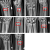
Example studies featuring the most common false negative cases. Lateral projections, if available, have been added to the figure to provide evidence for the presence of the fracture, but have not been part of the test set. (A) Distal radius fracture (red boxes) missed by all CNN models, but seen by 3 of 7 human raters. (B) Subtle dorsal distal radius fracture, missed by all CNN models and human experts. (C–E) Incomplete distal radius fractures missed by all raters but detected by the three CNN models.
Figure 6
Figure 6
Example studies featuring the most common false positive cases. (A) EfficientNet-B4 and ResNet-152 predicted a fracture in a case categorized as negative by the reference radiologists. There is subtlest buckling next to the asterisk (*), so a fracture cannot be ultimately ruled out. (B) VGG16 and 3 of 7 human experts predicted a fracture in this asymptomatic patient's cropped hand x-ray (these studies do not include lateral projections). (C) A negative case rated positive for fracture by 3 of 7 human experts. The PA projection on the right was taken two years later. The arrows point at cortical irregularities commonly encountered in the pediatric distal radius, which are atypical for a fracture. The plus symbol (+) marks a discrete thickening of the distal radius, which is not moving away from the growth plate in the follow-up on the right. Therefore, a healed fracture is also unlikely in that location.
Figure 7
Figure 7
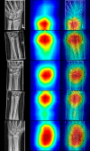
Random selection of radiographs positive for fracture with resNet-152 class activation maps (CAMs). The left column displays the source images, the middle column illustrates CAMs, and the right depicts fusions of source and sample images. Note that the classes are activated in the distal forearm region, even though the fields of view differ in the presented radiographs.

References

    1. Choi JW, Cho YJ, Lee S, Lee J, Lee S, Choi YH, et al. Using a dual-input convolutional neural network for automated detection of pediatric supracondylar fracture on conventional radiography. Invest Radiol. (2020) 55:101–10. 10.1097/RLI.0000000000000615 - DOI - PubMed
    1. Chung SW, Han SS, Lee JW, Oh KS, Kim NR, Yoon JP, et al. Automated detection and classification of the proximal humerus fracture by using deep learning algorithm. Acta Orthop. (2018) 89:468–73. 10.1080/17453674.2018.1453714 - DOI - PMC - PubMed
    1. Lindsey R, Daluiski A, Chopra S, Lachapelle A, Mozer M, Sicular S, et al. Deep neural network improves fracture detection by clinicians. Proc Natl Acad Sci U S A. (2018) 115:11591–6. 10.1073/pnas.1806905115 - DOI - PMC - PubMed
    1. LeCun Y, Bengio Y, Hinton G. Deep learning. Nature. (2015) 521:436–44. 10.1038/nature14539 - DOI - PubMed
    1. Bengio Y, Courville A, Vincent P. Representation learning: a review and new perspectives. IEEE Trans Pattern Anal Mach Intell. (2013) 35:1798–828. 10.1109/TPAMI.2013.50 - DOI - PubMed