

Identification and mechanistic basis of non-ACE2 blocking neutralizing antibodies from COVID-19 patients with deep RNA sequencing and molecular dynamics simulations
- PMID: 36589229
- PMCID: PMC9800910
- DOI: 10.3389/fmolb.2022.1080964
Identification and mechanistic basis of non-ACE2 blocking neutralizing antibodies from COVID-19 patients with deep RNA sequencing and molecular dynamics simulations
Abstract
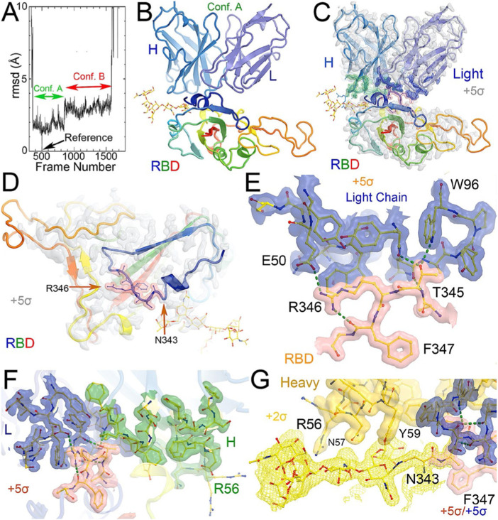
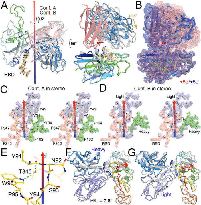
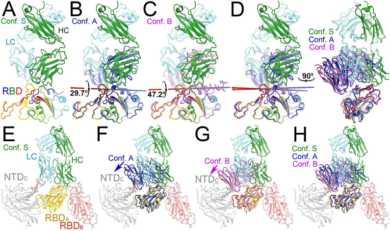
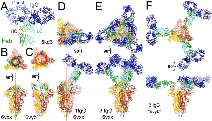
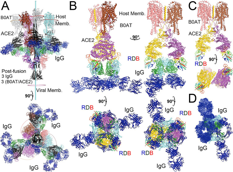
Variants of severe acute respiratory syndrome coronavirus-2 (SARS-CoV-2) continue to cause disease and impair the effectiveness of treatments. The therapeutic potential of convergent neutralizing antibodies (NAbs) from fully recovered patients has been explored in several early stages of novel drugs. Here, we identified initially elicited NAbs (Ig Heavy, Ig lambda, Ig kappa) in response to COVID-19 infection in patients admitted to the intensive care unit at a single center with deep RNA sequencing (>100 million reads) of peripheral blood as a diagnostic tool for predicting the severity of the disease and as a means to pinpoint specific compensatory NAb treatments. Clinical data were prospectively collected at multiple time points during ICU admission, and amino acid sequences for the NAb CDR3 segments were identified. Patients who survived severe COVID-19 had significantly more of a Class 3 antibody (C135) to SARS-CoV-2 compared to non-survivors (15059.4 vs. 1412.7, p = 0.016). In addition to highlighting the utility of RNA sequencing in revealing unique NAb profiles in COVID-19 patients with different outcomes, we provided a physical basis for our findings via atomistic modeling combined with molecular dynamics simulations. We established the interactions of the Class 3 NAb C135 with the SARS-CoV-2 spike protein, proposing a mechanistic basis for inhibition via multiple conformations that can effectively prevent ACE2 from binding to the spike protein, despite C135 not directly blocking the ACE2 binding motif. Overall, we demonstrate that deep RNA sequencing combined with structural modeling offers the new potential to identify and understand novel therapeutic(s) NAbs in individuals lacking certain immune responses due to their poor endogenous production. Our results suggest a possible window of opportunity for administration of such NAbs when their full sequence becomes available. A method involving rapid deep RNA sequencing of patients infected with SARS-CoV-2 or its variants at the earliest infection time could help to develop personalized treatments using the identified specific NAbs.
Keywords: COVID-19; RNA sequencing; antibodies; intensive care; sepsis.
Copyright © 2022 Fredericks, East, Shi, Liu, Maschietto, Ayala, Cioffi, Cohen, Fairbrother, Lefort, Nau, Levy, Wang, Batista, Lisi and Monaghan.
Conflict of interest statement
The authors declare that the research was conducted in the absence of any commercial or financial relationships that could be construed as a potential conflict of interest.
Figures
Similar articles
-
Effects of human anti-spike protein receptor binding domain antibodies on severe acute respiratory syndrome coronavirus neutralization escape and fitness.J Virol. 2014 Dec;88(23):13769-80. doi: 10.1128/JVI.02232-14. Epub 2014 Sep 17. J Virol. 2014. PMID: 25231316 Free PMC article.
-
Evaluating the Association of Clinical Characteristics With Neutralizing Antibody Levels in Patients Who Have Recovered From Mild COVID-19 in Shanghai, China.JAMA Intern Med. 2020 Oct 1;180(10):1356-1362. doi: 10.1001/jamainternmed.2020.4616. JAMA Intern Med. 2020. PMID: 32808970 Free PMC article.
-
Epitope Classification and RBD Binding Properties of Neutralizing Antibodies Against SARS-CoV-2 Variants of Concern.Front Immunol. 2021 Jun 4;12:691715. doi: 10.3389/fimmu.2021.691715. eCollection 2021. Front Immunol. 2021. PMID: 34149735 Free PMC article.
-
SARS-CoV-2 spike S2-specific neutralizing antibodies.Emerg Microbes Infect. 2023 Dec;12(2):2220582. doi: 10.1080/22221751.2023.2220582. Emerg Microbes Infect. 2023. PMID: 37254830 Free PMC article. Review.
-
Structural and antigenic variations in the spike protein of emerging SARS-CoV-2 variants.PLoS Pathog. 2022 Feb 17;18(2):e1010260. doi: 10.1371/journal.ppat.1010260. eCollection 2022 Feb. PLoS Pathog. 2022. PMID: 35176090 Free PMC article. Review.
Cited by
-
COVID-19 in patients with anemia and haematological malignancies: risk factors, clinical guidelines, and emerging therapeutic approaches.Cell Commun Signal. 2024 Feb 15;22(1):126. doi: 10.1186/s12964-023-01316-9. Cell Commun Signal. 2024. PMID: 38360719 Free PMC article. Review.
-
A computationally designed ACE2 decoy has broad efficacy against SARS-CoV-2 omicron variants and related viruses in vitro and in vivo.Commun Biol. 2023 May 12;6(1):513. doi: 10.1038/s42003-023-04860-9. Commun Biol. 2023. PMID: 37173421 Free PMC article.
-
Optimizing the breadth of SARS-CoV-2-neutralizing antibodies in vivo and in silico.Hum Vaccin Immunother. 2025 Dec;21(1):2526873. doi: 10.1080/21645515.2025.2526873. Epub 2025 Jul 21. Hum Vaccin Immunother. 2025. PMID: 40690731 Free PMC article. Review.
References
-
- Acevedo M. L., Alonso-Palomares L., Bustamante A., Gaggero A., Paredes F., Cortés C. P., et al. (2021). Infectivity and immune escape of the new SARS-CoV-2 variant of interest Lambda. medRxiv. 10.1101/2021.06.28.21259673 - DOI
-
- Andrews S. (2014). A quality control tool for high throughput sequence data. FastQC. Available at: http://www.bioinformatics.babraham.ac.uk/projects/fastqc/ (Accessed June 16, 2020).
Grants and funding
LinkOut - more resources
Full Text Sources
Other Literature Sources
Miscellaneous