

Action potential variability in human pluripotent stem cell-derived cardiomyocytes obtained from healthy donors
- PMID: 36589430
- PMCID: PMC9800870
- DOI: 10.3389/fphys.2022.1077069
Action potential variability in human pluripotent stem cell-derived cardiomyocytes obtained from healthy donors
Abstract
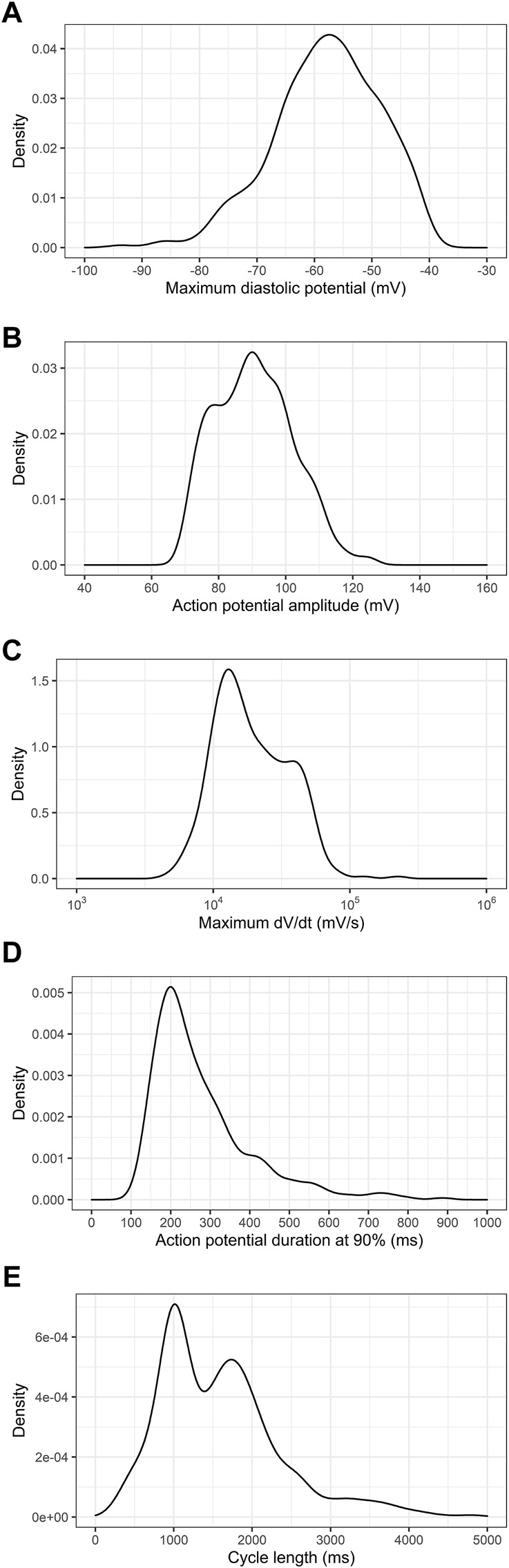
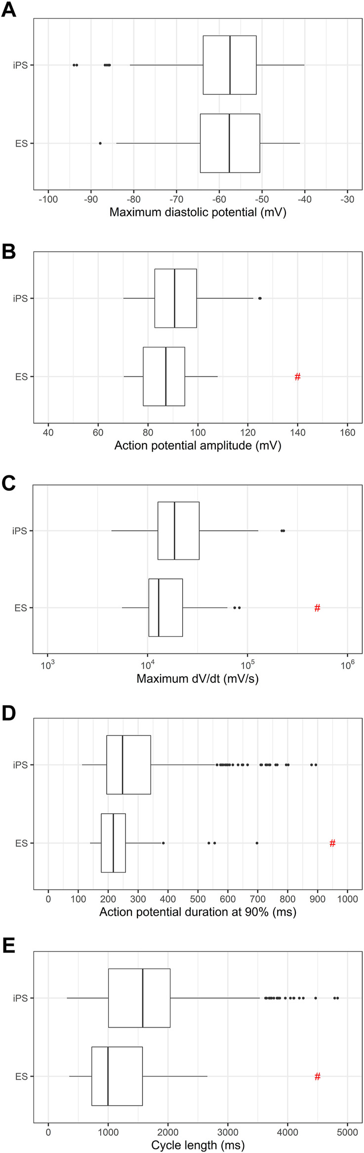
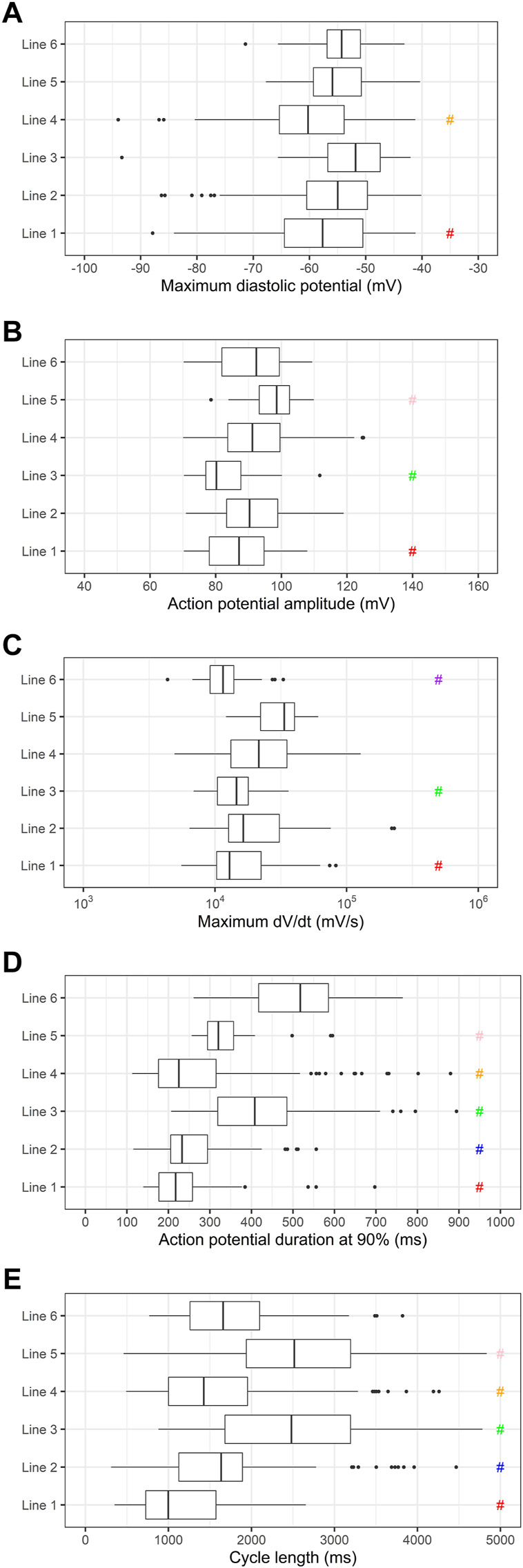
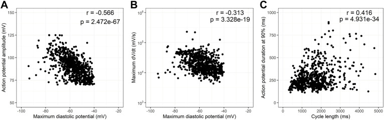
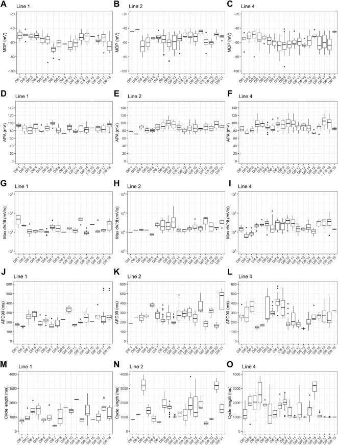
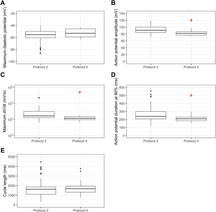
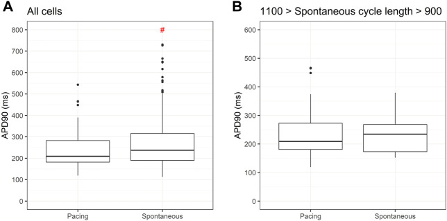
Human pluripotent stem cells (PSC) have been used for disease modelling, after differentiation into the desired cell type. Electrophysiologic properties of cardiomyocytes derived from pluripotent stem cells are extensively used to model cardiac arrhythmias, in cardiomyopathies and channelopathies. This requires strict control of the multiple variables that can influence the electrical properties of these cells. In this article, we report the action potential variability of 780 cardiomyocytes derived from pluripotent stem cells obtained from six healthy donors. We analyze the overall distribution of action potential (AP) data, the distribution of action potential data per cell line, per differentiation protocol and batch. This analysis indicates that even using the same cell line and differentiation protocol, the differentiation batch still affects the results. This variability has important implications in modeling arrhythmias and imputing pathogenicity to variants encountered in patients with arrhythmic diseases. We conclude that even when using isogenic cell lines to ascertain pathogenicity to variants associated to arrythmias one should use cardiomyocytes derived from pluripotent stem cells using the same differentiation protocol and batch and pace the cells or use only cells that have very similar spontaneous beat rates. Otherwise, one may find phenotypic variability that is not attributable to pathogenic variants.
Keywords: action potential (AP); cardiomyocytes; cell lines; differentiation batches; differentiation methods; healthy donors; iPSC (induced pluripotent stem cell); variability.
Copyright © 2022 Carvalho, Coutinho, Barbosa, Campos, Leitão, Pinto, Dos Santos, Farjun, De Araújo, Mesquita, Monnerat-Cahli, Medei, Kasai-Brunswick and De Carvalho.
Conflict of interest statement
The authors declare that the research was conducted in the absence of any commercial or financial relationships that could be construed as a potential conflict of interest.
Figures

Similar articles
-
Patient-Specific and Gene-Corrected Induced Pluripotent Stem Cell-Derived Cardiomyocytes Elucidate Single-Cell Phenotype of Short QT Syndrome.Circ Res. 2019 Jan 4;124(1):66-78. doi: 10.1161/CIRCRESAHA.118.313518. Circ Res. 2019. PMID: 30582453
-
Determining the Pathogenicity of a Genomic Variant of Uncertain Significance Using CRISPR/Cas9 and Human-Induced Pluripotent Stem Cells.Circulation. 2018 Dec 4;138(23):2666-2681. doi: 10.1161/CIRCULATIONAHA.117.032273. Circulation. 2018. PMID: 29914921 Free PMC article.
-
Evaluation of Batch Variations in Induced Pluripotent Stem Cell-Derived Human Cardiomyocytes from 2 Major Suppliers.Toxicol Sci. 2017 Mar 1;156(1):25-38. doi: 10.1093/toxsci/kfw235. Toxicol Sci. 2017. PMID: 28031415
-
Human Induced Pluripotent Stem Cell-Derived Cardiomyocytes as Models for Cardiac Channelopathies: A Primer for Non-Electrophysiologists.Circ Res. 2018 Jul 6;123(2):224-243. doi: 10.1161/CIRCRESAHA.118.311209. Circ Res. 2018. PMID: 29976690 Free PMC article. Review.
-
Addressing Variability and Heterogeneity of Induced Pluripotent Stem Cell-Derived Cardiomyocytes.Adv Exp Med Biol. 2020;1212:1-29. doi: 10.1007/5584_2019_350. Adv Exp Med Biol. 2020. PMID: 30850960 Review.
Cited by
-
Hydrogel-Sheathed hiPSC-Derived Heart Microtissue Enables Anchor-Free Contractile Force Measurement.Adv Sci (Weinh). 2023 Dec;10(35):e2301831. doi: 10.1002/advs.202301831. Epub 2023 Oct 17. Adv Sci (Weinh). 2023. PMID: 37849230 Free PMC article.
-
Revolutionizing Disease Modeling: The Emergence of Organoids in Cellular Systems.Cells. 2023 Mar 18;12(6):930. doi: 10.3390/cells12060930. Cells. 2023. PMID: 36980271 Free PMC article. Review.
-
Optical redox imaging to screen synthetic hydrogels for stem cell-derived cardiomyocyte differentiation and maturation.Biophotonics Discov. 2024 May;1(1):015002. doi: 10.1117/1.bios.1.1.015002. Epub 2024 May 20. Biophotonics Discov. 2024. PMID: 39036366 Free PMC article.
-
iPSC-Derived Biological Pacemaker-From Bench to Bedside.Cells. 2024 Dec 11;13(24):2045. doi: 10.3390/cells13242045. Cells. 2024. PMID: 39768137 Free PMC article. Review.
References
-
- Burridge P. W., Thompson S., Millrod M. A., Weinberg S., Yuan X., Peters A., et al. (2011). A universal system for highly efficient cardiac differentiation of human induced pluripotent stem cells that eliminates interline variability. Plos One 6, e18293. 10.1371/journal.pone.0018293 - DOI - PMC - PubMed
-
- Cruvinel E., Ogusuku I., Cerioni R., Rodrigues S., Gonçalves J., Góes M. E., et al. (2020). Long-term single-cell passaging of human iPSC fully supports pluripotency and high-efficient trilineage differentiation capacity. SAGE Open Med. 8, 2050312120966456. 10.1177/2050312120966456 - DOI - PMC - PubMed
LinkOut - more resources
Full Text Sources