

Appendiceal schwannoma presenting as acute appendicitis
- PMID: 36589492
- PMCID: PMC9794891
- DOI: 10.1016/j.radcr.2022.11.054
Appendiceal schwannoma presenting as acute appendicitis
Abstract
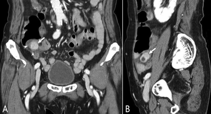
Schwannomas are nerve sheath tumors that rarely occur in the gastrointestinal tract. In the gastrointestinal tract, schwannomas are mostly found in the stomach and small bowel. Herein, we describe a case of appendiceal schwannoma that caused appendix obstruction and developed acute appendicitis. An 83-year-old woman was admitted to the emergency department with 3 days' history of abdominal pain. She had tenderness and rebound tenderness in the lower right quadrant. Computed tomography revealed a 1.3 cm mass in the appendix orifice, with associated distal appendiceal dilatation and wall thickening. The patient underwent emergency surgery (laparoscopic partial cecectomy). Histopathological examination confirmed that the mass was a schwannoma and was associated with acute suppurative appendicitis. Our case is significant in that it adds to another rarely reported case of appendiceal schwannoma. Moreover, it is important to recognize the presence of an appendiceal tumor associated with acute appendicitis.
Keywords: Appendiceal schwannoma; Appendicitis; Case report; Computed tomography.
© 2022 The Authors. Published by Elsevier Inc. on behalf of University of Washington.
Figures


References
-
- Kang DB, Kim SH, Oh JT, Park WC, Lee JK, Kim HS. Colonic schwannoma. J Korean Surg Soc. 2007:183–187.
Publication types
LinkOut - more resources
Full Text Sources
