

Hydatid cyst of the uterus: Very rare location
- PMID: 36589497
- PMCID: PMC9798129
- DOI: 10.1016/j.radcr.2022.10.068
Hydatid cyst of the uterus: Very rare location
Abstract
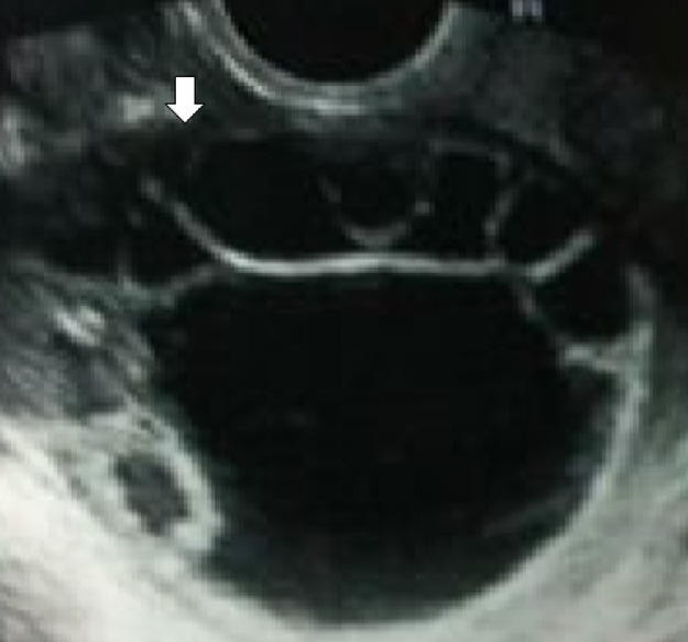
The involvement of the genital tract of a hydatid cyst is rare and the occurrence in the uterus is an extreme rarity. The diagnosis of this localization is difficult because the clinical and radiological findings are often misleading and the diagnosis is often worn during surgery and after histopathological examination of the surgical specimen. We report the case of a patient who consulted for primary infertility, with a clinical finding as the only anomaly significantly large uterus, and imaging pointing strongly toward an ovarian multilocular cyst, and in which the discovery of hydatid cyst was accidental intraoperative with double localization uterine and omental. Radical treatment cannot be discussed in this young patient of 32 years and gravid 0. The removal of the cyst wall completely and excision of the mass epiploic seemed reasonable. The patient was placed under Mebendazol and is always under the supervision of a possible recurrence.
Keywords: Hydatid cyst; Omentum; Pelvic; Uterus.
© 2022 The Authors. Published by Elsevier Inc. on behalf of University of Washington.
Figures


References
-
- Boufettal R., Lefryiekh M.R., Fadil A., Ouariti Zerouali N. Kyste Hydatique Pelvien Primitif (A Propos D'un Cas) J Maroc Urol. 2008;9:34–36.
-
- Arslan H, Sakarya M, Bozkurt M, Dilek F, Yilmaz Y, Dilek O, et al. Free hydatid cyst only covered with germinative membrane disrupted from fibrotic capsule in the peritoneal cavity: a case report. Acta Chir. 1998;9:85–86. - PubMed
-
- Alami A, Bouftila H, Essaadi F, et al. Hydatidose Peritoneale A Propos D’1 Cas. Med Maghreb. 2006;135:35–37.
Publication types
LinkOut - more resources
Full Text Sources
