. 2022 Dec 23:2022:8704016.
doi: 10.1155/2022/8704016. eCollection 2022.

Inhibition of PFKFB Preserves Intestinal Barrier Function in Sepsis by Inhibiting NLRP3/GSDMD

Affiliations

Inhibition of PFKFB Preserves Intestinal Barrier Function in Sepsis by Inhibiting NLRP3/GSDMD

Yongsheng Zhang et al. Oxid Med Cell Longev. .

Abstract

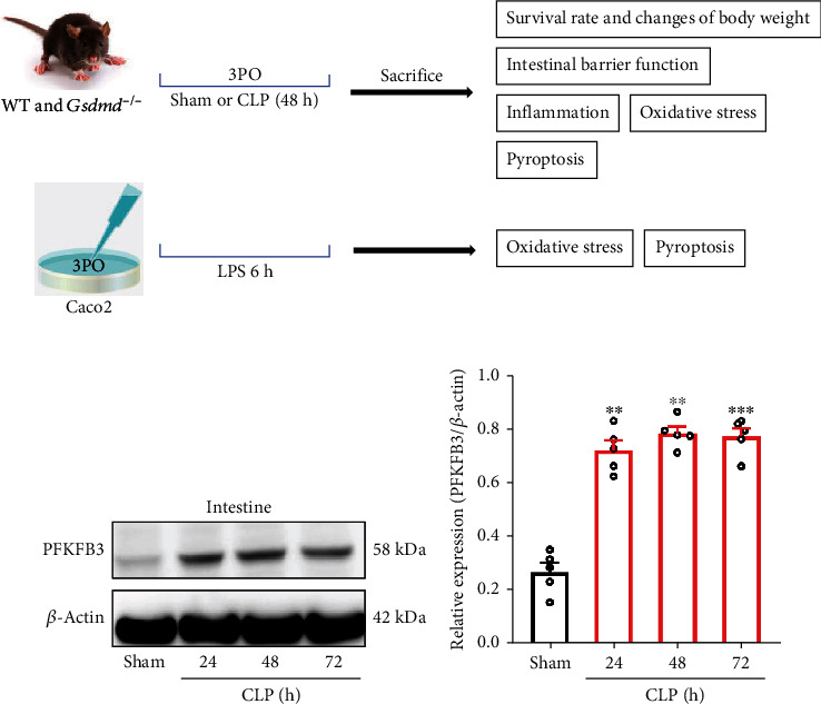
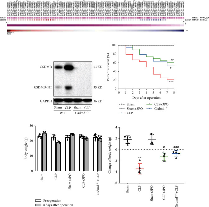
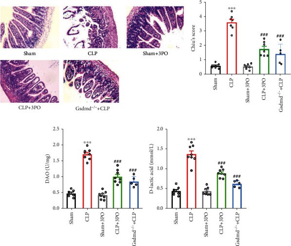
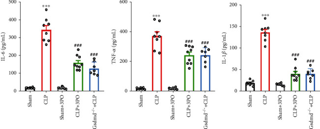
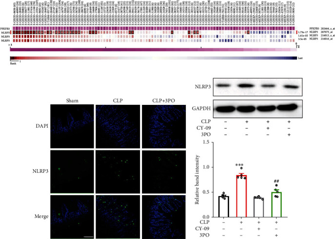
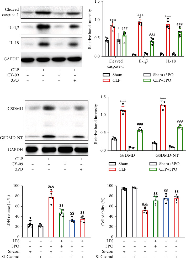
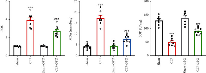
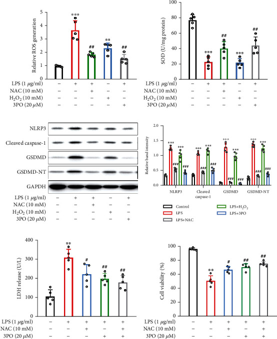
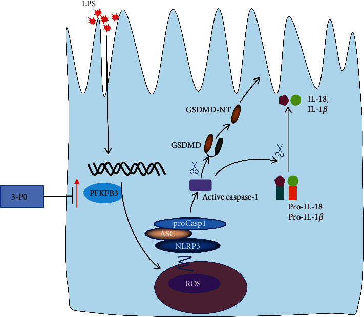
Intestinal barrier dysfunction is associated with the occurrence and development of sepsis. Further, aerobic glycolysis plays an essential role in inflammation and cell death. This study is aimed at investigating the protective effect and mechanism of PFKFB3 inhibition on intestinal barrier dysfunction in sepsis mice. Sepsis mouse models were established by cecal ligation and puncture (CLP) in wild-type mice and Gsdmd-/- mice. The results showed that the expression of 6-phosphofructo-2-kinase/fructose-2,6-biphosphatase 3 (PFKFB3) in the small intestines was significantly upregulated in sepsis. 3-(3-Pyridinyl)-1-(4-pyridinyl)-2-propen-1-one (3PO), the specific inhibitor of PFKFB3, and Gsdmd gene knockout significantly inhibited the inflammatory response and cell death caused by sepsis, thus alleviating intestinal damage and barrier dysfunction. 3PO was also shown to significantly inhibit oxidative stress and NLRP3/caspase-1/GSDMD-dependent cell pyroptosis in the small intestines. The in vitro studies revealed that 3PO reduced NLRP3/caspase-1/GSDMD-dependent cell pyroptosis by inhibiting ROS. Taken together, our results suggest that PFKFB3 is involved in inflammation, oxidative stress, and pyroptosis during sepsis and enhances intestinal damage, which may provide important clues about the potential targets to be exploited in this highly lethal disease.


Conflict of interest statement

The authors declare that the research was conducted in the absence of any commercial or financial relationships that could be construed as a potential conflict of interest.

Figures

Figure 1
Figure 1
The expression of PFKFB3 was elevated in the intestinal tissues of septic mice. (a) The experimental design of this study. (b, c) The expression of PFKFB3 was analyzed by Western blot at different time points after CLP. Experimental values are expressed as means ± SD (n = 5 per group). Statistical analysis was performed using one-way ANOVA (c). ∗∗P < 0.01 and ∗∗∗P < 0.001.
Figure 2
Figure 2
Inhibition of PFKFB3 and Gsdmd gene knockout can improve the prognosis of septic mice. (a) The coexpression of PFKFB3 and GSDMD was analyzed by MEM. (b, c) The LPS-induced PFKFB3 expression was inhibited by 3PO in Caco-2 cells. (d) GSDMD and GSDMD-NT expression in intestinal tissues of wild-type and Gsdmd−/− mice after sham or CLP operation. (e) Survival rates were calculated in different groups. The values were shown as mean ± SD (n = 5 per group in (d) and (e)). Statistical analysis was performed using a t-test (e) and log-rank test (c). ∗∗∗P < 0.001 versus sham group; #P < 0.05, ##P < 0.01, and ###P < 0.001 versus CLP group.
Figure 3
Figure 3
Effects of 3PO or Gsdmd genetic deletion on intestinal histology and intestinal barrier function. At 24 h after sham or CLP operation, the histopathological alterations of gut tissues were measured using H-E staining (a) and analyzed by Chiu's score (b). Scale bar = 100 μm. (c) Detection of DAO and (d) D-lactic acid levels in serum. The values were shown as mean ± SD (n = 7 per group). Statistical analysis was performed using a t-test (b–d). ∗∗∗P < 0.001 versus sham group; ###P < 0.001 versus CLP group.
Figure 4
Figure 4
Effect of 3PO and Gsdmd gene knockout on inflammatory cytokines in intestinal tissue. (a–c) Levels of IL-6, TNF-α, and IL-1β in small intestinal tissues were measured by ELISA. The values were shown as mean ± SD (n = 6 − 9). ∗∗∗P < 0.001 versus sham group; ###P < 0.001 versus CLP group. The values were shown as mean ± SD (n = 5). Statistical analysis was performed using a t-test (a–c). ∗∗∗P < 0.001 compared with the no-treatment group and ##P < 0.01 compared with the CLP treatment group.
Figure 5
Figure 5
3PO downregulates NLRP3 expression. The coexpression of PFKFB3 and NLRP3 was analyzed by MEM (a). The expression of NLRP3 was detected in small intestinal tissues by immunofluorescence (b) and Western blot (c). The values were shown as mean ± SD (n = 5). Statistical analysis was performed using a t-test (c). ∗∗∗P < 0.001 compared with the no-treatment group and ##P < 0.01 compared with the CLP treatment group.
Figure 6
Figure 6
3PO protects against pyroptosis in vitro and in vivo. (a, b) Pyroptosis-related proteins, including NLRP3, cleaved caspase-1, IL-1β, and IL-18, were examined by Western blot in intestinal tissue of mice. (c, d) The expression of GSDMD was detected by Western blot in intestinal tissue of mice. si-Gsdmd was used to inhibit pyroptosis and reveal the relationship between 3PO and pyroptosis. (e) Levels of the released LDH were detected in C2C12 culture medium supernatant. (f) Cell viability was determined using the CCK-8 assay. The values were shown as mean ± SD (n = 5). Statistical analysis was performed using a t-test (b, d–f). ∗∗∗P < 0.001 compared with the no-treatment group and ###P < 0.001 compared with the CLP treatment group. &&P < 0.01 compared with the si-Con group and $$P < 0.01 compared with the LPS+si-Con group. 3PO protected against CLP-induced intestinal injury by suppressing NLRP3/caspase-1/GSDMD.
Figure 7
Figure 7
3PO alleviates oxidative stress in intestinal tissues. (a–c) Oxidative stress was measured by ROS, MDA, and SOD. The values were shown as mean ± SD (n = 7 − 9). Statistical analysis was performed using a t-test (a–c). ∗∗∗P < 0.001 compared with the sham group and ###P < 0.001 compared with the CLP treatment group.
Figure 8
Figure 8
3PO suppressed pyroptosis by inhibiting ROS. (a, b) Oxidative stress was measured by ROS and SOD in vitro. (c, d) Pyroptosis-related proteins were detected by Western blot. (e) The released levels of LDH were detected. (f) Cell viability was measured using the CCK-8 assay. The values were shown as mean ± SD (n = 5). Statistical analysis was performed using a t-test (a, b, d–f). ∗∗∗P < 0.001 compared with the no-treatment group and ###P < 0.001 compared with the LPS treatment group.
Figure 9
Figure 9
Schematic model for the inhibition of PFKFB3 preserves intestinal barrier function in sepsis via preventing GSDMD-dependent pyroptosis by inhibiting ROS production.

Similar articles

Cited by

References

    1. Gheen N. Sepsis-3 definitions. Annals of Emergency Medicine . 2016;68(6):784–785. doi: 10.1016/j.annemergmed.2016.07.008. - DOI - PubMed
    1. Meng M., Klingensmith N. J., Coopersmith C. M. New insights into the gut as the driver of critical illness and organ failure. Current Opinion in Critical Care . 2017;23(2):143–148. doi: 10.1097/MCC.0000000000000386. - DOI - PMC - PubMed
    1. Quan R., Chen C., Yan W., Zhang Y., Zhao X., Fu Y. BAFF blockade attenuates inflammatory responses and intestinal barrier dysfunction in a murine endotoxemia model. Frontiers in Immunology . 2020;11, article 570920 doi: 10.3389/fimmu.2020.570920. - DOI - PMC - PubMed
    1. Haussner F., Chakraborty S., Halbgebauer R., Huber-Lang M. Challenge to the intestinal mucosa during sepsis. Frontiers in Immunology . 2019;10:p. 891. doi: 10.3389/fimmu.2019.00891. - DOI - PMC - PubMed
    1. Tan C., Gu J., Li T., et al. Inhibition of aerobic glycolysis alleviates sepsis‑induced acute kidney injury by promoting lactate/sirtuin 3/AMPK‑regulated autophagy. International Journal of Molecular Medicine . 2021;47(3) doi: 10.3892/ijmm.2021.4852. - DOI - PMC - PubMed

Substances