. 2022 Jul 13;11(6):540-557.
doi: 10.1159/000525375. eCollection 2022 Dec.

Sulfatase-2 from Cancer Associated Fibroblasts: An Environmental Target for Hepatocellular Carcinoma?

Affiliations

Sulfatase-2 from Cancer Associated Fibroblasts: An Environmental Target for Hepatocellular Carcinoma?

Marco Y W Zaki et al. Liver Cancer. .

Abstract

Introduction: Heparin sulphate proteoglycans in the liver tumour microenvironment (TME) are key regulators of cell signalling, modulated by sulfatase-2 (SULF2). SULF2 overexpression occurs in hepatocellular carcinoma (HCC). Our aims were to define the nature and impact of SULF2 in the HCC TME.

Methods: In liver biopsies from 60 patients with HCC, expression and localization of SULF2 were analysed associated with clinical parameters and outcome. Functional and mechanistic impacts were assessed with immunohistochemistry (IHC), in silico using The Cancer Genome Atlas (TGCA), in primary isolated cancer activated fibroblasts, in monocultures, in 3D spheroids, and in an independent cohort of 20 patients referred for sorafenib. IHC targets included αSMA, glypican-3, β-catenin, RelA-P-ser536, CD4, CD8, CD66b, CD45, CD68, and CD163. SULF2 impact of peripheral blood mononuclear cells was assessed by migration assays, with characterization of immune cell phenotype using fluorescent activated cell sorting.

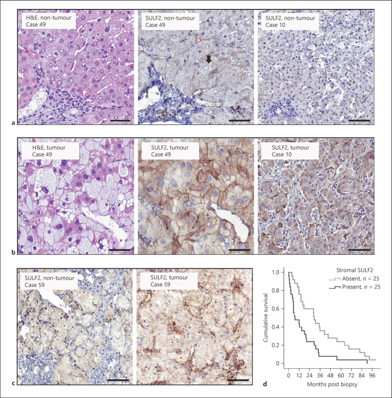
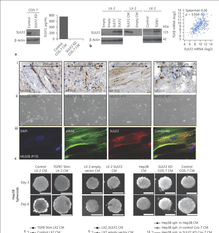
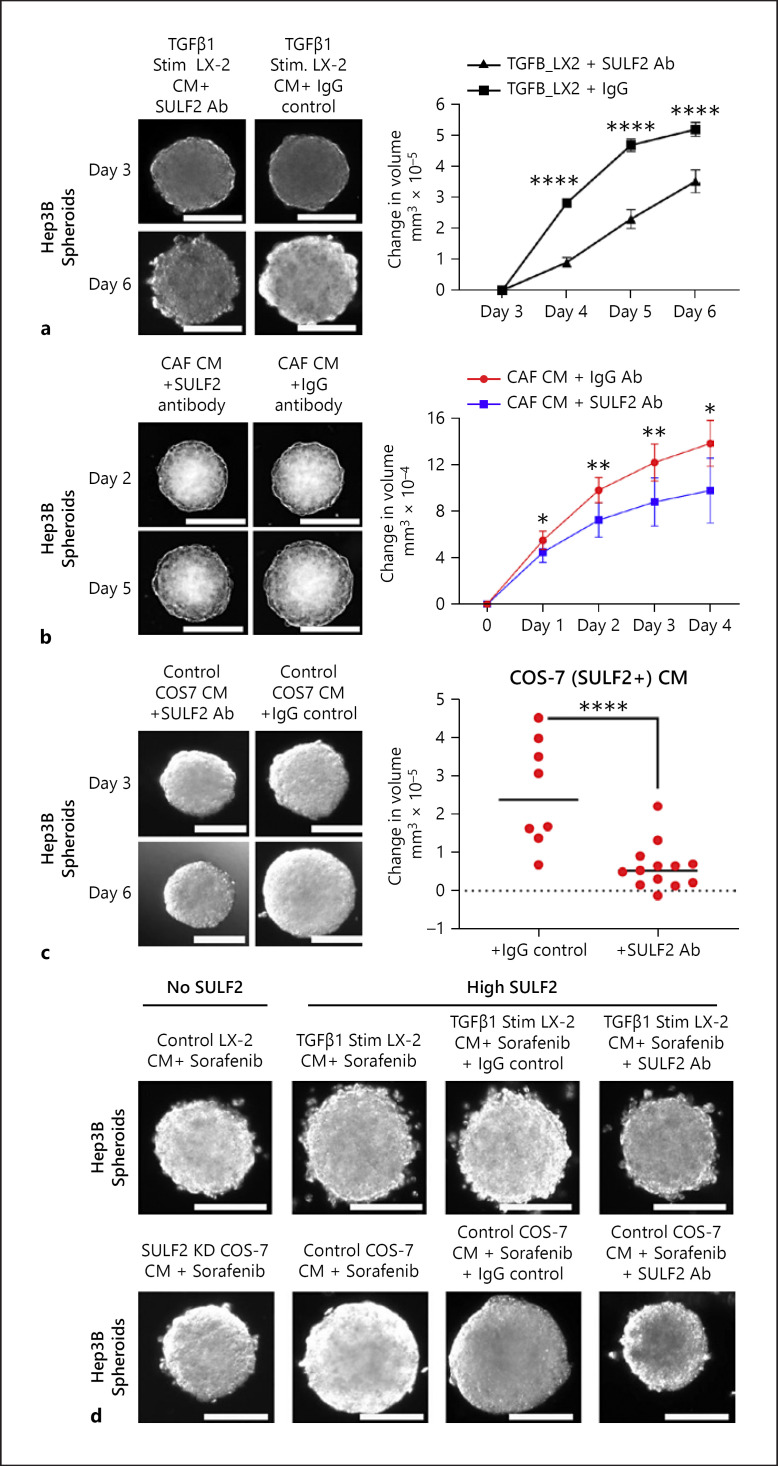
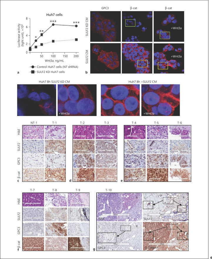
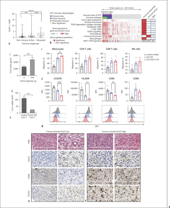
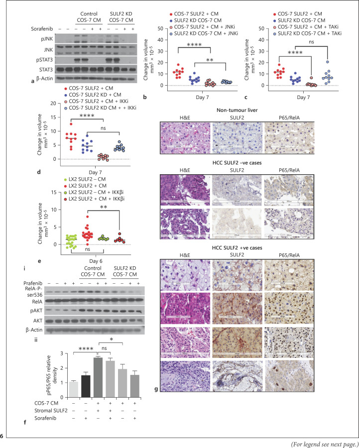
Results: We report that while SULF2 was expressed in tumour cells in 15% (9/60) of cases, associated with advanced tumour stage and type 2 diabetes, SULF2 was more commonly expressed in cancer-associated fibroblasts (CAFs) (52%) and independently associated with shorter survival (7.2 vs. 29.2 months, p = 0.003). Stromal SULF2 modulated glypican-3/β-catenin signalling in vitro, although in vivo associations suggested additional mechanisms underlying the CAF-SULF2 impact on prognosis. Stromal SULF2 was released by CAFS isolated from human HCC. It was induced by TGFβ1, promoted HCC proliferation and sorafenib resistance, with CAF-SULF2 linked to TGFβ1 and immune exhaustion in TGCA HCC patients. Autocrine activation of PDGFRβ/STAT3 signalling was evident in stromal cells, with the release of the potent monocyte/macrophage chemoattractant CCL2 in vitro. In human PBMCs, SULF2 preferentially induced the migration of macrophage precursors (monocytes), inducing a phenotypic change consistent with immune exhaustion. In human HCC tissues, CAF-SULF2 was associated with increased macrophage recruitment, with tumouroid studies showing stromal-derived SULF2-induced paracrine activation of the IKKβ/NF-κB pathway, tumour cell proliferation, invasion, and sorafenib resistance.

Conclusion: SULF2 derived from CAFs modulates glypican-3/β-catenin signalling but also the HCC immune TME, associated with tumour progression and therapy resistance via activation of the TAK1/IKKβ/NF-κB pathway. It is an attractive target for combination therapies for patients with HCC.

Keywords: Biomarkers; Glypican-3; Hepatocellular carcinoma; Inflammation; Metabolic disease; Molecular targets; NASH; Spheroids; Sulfatase-2; Tumour microenvironment.


Conflict of interest statement

F.O. is a director and shareholder in Fibrofind Ltd. J.L. is a shareholder in Fibrofind Ltd, all other authors declare no competing interests.

Figures

Fig. 1
Fig. 1
SULF2 expression in non-tumour liver and HCC tumour cells predicts prognosis: Representative images show H&E staining and SULF2 IHC in non-tumour (a) and tumour paired tissues (b). In non-tumour liver, SULF2 was expressed on the canalicular surface hepatocytes (black arrow), hepatic arterioles (a), endothelial cells (e), and occasional nonparenchymal sinusoidal cells (white arrows). In the tumour, cytoplasmic (right image) and membranous SULF2 (middle image) was increased in neoplastic cells. c Images show SULF2 IHC in non-tumour and tumour paired tissue. SULF2 was upregulated in CAFs in tumour tissue as compared to minimal expression in non-tumour stroma. Images are ×20 magnification and scale bars represent 50 microns. d Survival of patients with stromal SULF2 present in their tumour was markedly reduced (median 7.2 months) compared to patients in whom it was absent/scanty (median 29.2 months; Kaplan-Meier, p = 0.003).
Fig. 2
Fig. 2
SULF2 is expressed by CAFS isolated from SULF2 stromal positive tumours, with SULF2 conditioned media stimulating growth of 3D Hep3B spheroids in vitro. a SULF2 was highly expressed in COS-7 cells and suppressed by SULF2 shRNA, assessed by Western blot and ELISA assay. b Western blot of LX-2 whole cell lysates showing LX-2 cells with little SULF2 (left panel). SULF2 expression was induced in LX-2 after transfection with a SULF2 expression vector compared to empty vector control (left). SULF2 levels were elevated in CM from LX-2 cells transfected with a SULF2 expression vector (middle). TGFβ stimulation of LX-2 cells induced SULF2 expression, with a highly significant correlation between mRNA of the two in TCGA dataset (right panels). Mixed cell isolations from SULF2 stromal positive tumours (ci) yielded CAFs after trypsinisation and replating in fibroblast culture media (cii). ciiiDual labelling immunofluorescence confirmed co-expression of SULF2 (red) and αSMA (green) in primary CAFs. CM from TGFβ-stimulated (d) or SULF2 expression vector-transfected LX-2 cells (e) promoted growth of Hep3B (SULF2 null) spheroids. f CM from control COS-7 cells expressing SULF2 promoted spheroid growth in Hep3B cells, as compared to culture with in SULF2 KD COS-7 CM. Scale bars represent 200 microns. Change in spheroid volume is in online supplementary Table 4. Data mean ± SEM; n = 7 to 10 spheroids per condition. **p < 0.01; ***p = 0.001; ****p < 0.001. “P” denotes number of passages for CAFs.
Fig. 3
Fig. 3
The impact of stromal SULF2 inhibition on the growth of tumour spheroids and SULF2-mediated sorafenib sensitivity: ac TGFβ-stimulated LX-2-CM (a), primary CAF-SULF2-CM (n = 4 SULF2-positive HCC) (b), and COS-7 CM (c) induced Hep3B spheroid growth was abrogated using SULF2 antibody versus control IgG. d Sorafenib treatment had little impact on the growth of Hep3B spheroids cultured in control TGFβ-stimulated LX-2 cell CM (upper images) or COS-7 cell CM (lower images). Growth was dramatically suppressed and sensitivity to sorafenib restored in Hep3B spheroids cultured CM from TGFβ-stimulated LX-2 or SULF2 KD COS-7 cells respectively. Changes in spheroid volume are in online supplementary Table 4. Data mean ± SEM; 7 to 10 spheroids per condition. ****p < 0.001.
Fig. 4
Fig. 4
SULF2, Glypican-3 and β-catenin in HCC. In HuH7 cells which overexpress SULF2 (a), addition of Wnt3a ligand stimulated a TOPFLASH β-catenin (β-cat) reporter, abrogated by SULF2 KD. b Confocal immunofluorescence staining of HuH7 cells showed low levels of glypican-3 (GPC3) and β-catenin in the presence of CM from COS-7 KD cells, +/− Wnt3a 100 ng/mL. In the presence of SULF2-CM, GPC3 and membranous β-cat were upregulated. There was evidence of nuclear β-cat in the presence of Wnt3a (magnified images). cg Haematoxylin and eosin images, with SULF2, glypican-3 and β-catenin immunohistochemistry is shown for 10 cases. A non-tumour (NT) case is in (c), with the corresponding tumour (T) showing tumour cells SULF2+, nonspecific nuclear GPC3, and nuclear β-cat. Panel (d) shows two SULF2+ HCC with grade 1–2 GPC3+ and nuclear β-cat. In (e), three CAF-SULF2+ cases show grade 1–2 GPC+ and nuclear β-cat. T7 (f), however, has no SULF2 or GCP3, but nuclear β-cat++; T8 has scant SULF2, grade 2 GPC3 and no nuclear β-cat, while T-9 is CAF-SULF2+, GPC3−, and β-cat++. T10 is from a resection, showing tumour heterogeneity, with mild tumour SULF2+ (right [R]), with nuclear β-cat, while a region with CAF-SULF2+ (left [L]) shows membranous accumulation of β-cat. GPC3+ was classed negative, with absent cytoplasmic or membranous stain in both areas.
Fig. 5
Fig. 5
SULF2 expression characterizes an immune HCC phenotype, with stromal SULF2 upregulating CCL2 inducing, an altered macrophage phenotype and accumulation of macrophage in tumours. a SULF2 expression in HCC patients with an immune-exhausted phenotype was higher than patients with resting and immune active phenotypes (p< 0.001, independent sample Kruskal-Wallis test, n = 370 patients). b Heatmap of TCGA patients classified as Immune (exhausted and active) or nonimmune (rest) illustrates further how SULF2 expression correlates with TGFβ1 signalling, TGFβ1 regulated signatures (TBRS), and immune signatures. The median of the enrichment score in each class is shown on the right, with the percentage of patients in each class with enrichment of the WNT-TGFB signature. An ELISA assay confirmed SULF2 overexpression in LX-2 CM, with promoted secretion of CCL2 shown in (ci). ciiCCL2 mRNA was suppressed in SULF2 KD COS-7 cells. diLX-2 SULF2-CM preferentially increased transwell migration of monocytes (macrophage precursors) relative to LX-2-CM. diiCharacterization of CM activated CD80 expressing monocytes revealed an altered phenotype, with reduced expression of CX3CR1, HLADR, and CD86 in the presence of SULF2. Representative images of H&E, SULF2, CD68, and CD163 immunohistochemistry in patients with low (e) versus high (f) tumour stromal SULF2 expression showed higher numbers of CD68 or CD163 positive macrophages in tumours with high SULF2.
Fig. 6
Fig. 6
Stromal SULF2-dependent Hep3B growth and sorafenib sensitivity were regulated by TAK1/IKKβ/NF-κB RelA-P-ser536, with nuclear p65 associated with stromal SULF2. a Western blotting of Hep3B whole cell lysates, showed that activation of JNK/pJNK and STAT3/pSTAT3 pathways in Hep3B cells was driven by COS-7 CM independently of SULF2 and was sensitive to sorafenib treatment. b JNK inhibition limited Hep3B spheroid growth in both SULF2-dependent and independent fashion. In contrast, TAK1 (c) or IKKβ (d) inhibition limited SULF2-dependant Hep3B spheroid growth. e Similarly, Hep3B spheroid growth promoted by CM from SULF2-expressing LX-2 cells was suppressed by IKKβ inhibition. fiActivation of RelA-P-ser536 in Hep3B cells was dependent on control COS-7 CM and persistent in the presence of sorafenib, as was AKT activation. Quantification of RelA-P-ser536 (p-p65) relative to total p65 detected by Western blot is shown in fii, where data are mean ± SEM of 4 experiments. g IHC staining confirmed that SULF2 positivity in stromal cells associated with nuclear RelA-P-ser536 in adjacent tumour cells. Images are at ×20 magnification; scale bars represent 100–200 microns. Hep3B spheroid data are expressed as mean ± SEM of n = 10 experimental repeats. ns, not significant, *p = 0.05,**p < 0.01; ***p < 0.001, ****p< 0.0001.

References

    1. Bray F, Ferlay J, Soerjomataram I, Siegel RL, Torre LA, Jemal A. Global cancer statistics 2018: GLOBOCAN estimates of incidence and mortality worldwide for 36 cancers in 185 countries. CA Cancer J Clin. 2018;68((6)):394–424. - PubMed
    1. Forner A, Reig M, Bruix J. Hepatocellular carcinoma. Lancet. 2018;391((10127)):1301–14. - PubMed
    1. Reeves HL, Zaki MYW, Day CP. Hepatocellular carcinoma in obesity, type 2 diabetes, and NAFLD. Dig Dis Sci. 2016;61((5)):1234–45. - PubMed
    1. Pinter M, Jain RK, Duda DG. The current landscape of immune checkpoint blockade in hepatocellular carcinoma: a review. JAMA Oncol. 2021;7((1)):113–23. - PMC - PubMed
    1. Morimoto-Tomita M, Uchimura K, Werb Z, Hemmerich S, Rosen SD. Cloning and characterization of two extracellular heparin-degrading endosulfatases in mice and humans. J Biol Chem. 2002;277((51)):49175–85. - PMC - PubMed