. 2022 Dec 14:10:1000553.
doi: 10.3389/fcell.2022.1000553. eCollection 2022.

A new model for fatty acid hydroxylase-associated neurodegeneration reveals mitochondrial and autophagy abnormalities

Affiliations

A new model for fatty acid hydroxylase-associated neurodegeneration reveals mitochondrial and autophagy abnormalities

Frida Mandik et al. Front Cell Dev Biol. .

Abstract

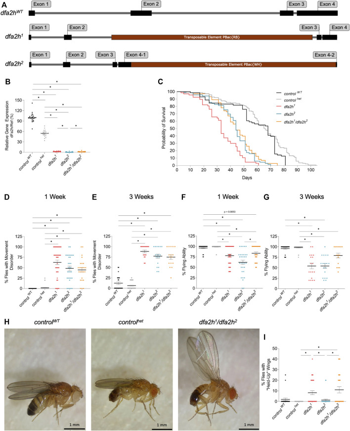
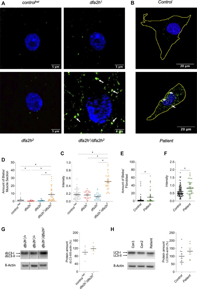
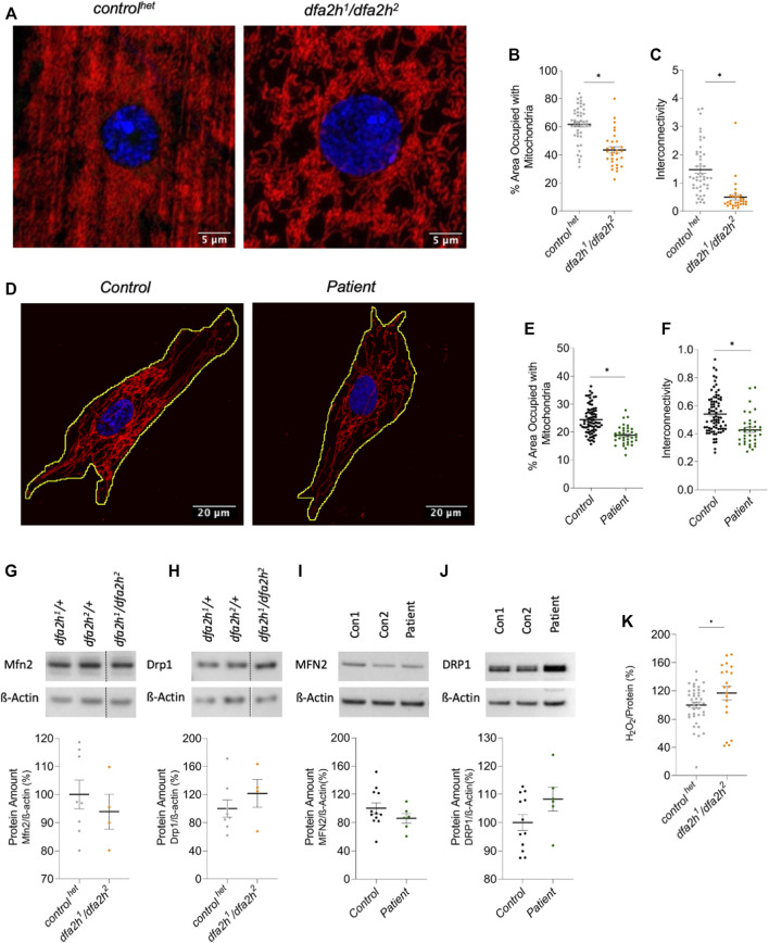
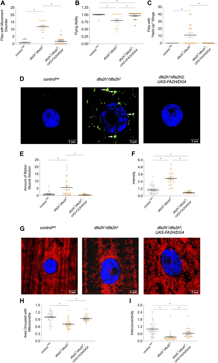
Fatty acid hydroxylase-associated neurodegeneration (FAHN) is a rare disease that exhibits brain modifications and motor dysfunctions in early childhood. The condition is caused by a homozygous or compound heterozygous mutation in fatty acid 2 hydroxylase (FA2H), whose encoded protein synthesizes 2-hydroxysphingolipids and 2-hydroxyglycosphingolipids and is therefore involved in sphingolipid metabolism. A few FAHN model organisms have already been established and give the first insight into symptomatic effects. However, they fail to establish the underlying cellular mechanism of FAHN so far. Drosophila is an excellent model for many neurodegenerative disorders; hence, here, we have characterized and validated the first FAHN Drosophila model. The investigation of loss of dfa2h lines revealed behavioral abnormalities, including motor impairment and flying disability, in addition to a shortened lifespan. Furthermore, alterations in mitochondrial dynamics, and autophagy were identified. Analyses of patient-derived fibroblasts, and rescue experiments with human FA2H, indicated that these defects are evolutionarily conserved. We thus present a FAHN Drosophila model organism that provides new insights into the cellular mechanism of FAHN.

Keywords: Drosophila melanogaster; FA2H; autophagy; fatty acid hydroxylase-associated neurodegeneration; mitochondria.


Conflict of interest statement

The authors declare that the research was conducted in the absence of any commercial or financial relationships that could be construed as a potential conflict of interest.

Figures

FIGURE 1
FIGURE 1
Transposable element insertion leads to reduced dfa2h cDNA level, causing reduced lifespan and motor disabilities. (A) Schematic image of the gDNA of dfa2h WT and the transposable element insertion lines dfa2h 1 and dfa2h 2 . (B) Relative dfa2h level normalized to the combined data of the reference genes Act5c, Rpl32, and eEF1α2 expressed as percentages relative to control WT that was set to 100%. The dots represent the single data points, and the lines show mean ± SEM (n = 21). (C) Survival monitoring of dfa2h mutant flies, control het and control WT (n = 67-137). Group curve comparisons were performed using log-Rank (Mantel-Cox) Test with a significance level of p < 0.05 (*). We performed a pairwise curve comparison using the log-Rank (Mantel-Cox) Test for better result interpretation. All pairwise comparisons, except the comparison of dfa2h 2 vs. dfa2h 1 /dfa2h 2 , were significant. (D) One-week-old and (E) three-week-old homozygous and compound heterozygous dfa2h flies display movement disabilities compared to control flies. (F) One-week-old and (G) three-week-old dfa2h-mutant flies show difficulties in flying capacity compared to control flies. (H) “Held-up” wing phenotype of dfa2h 1 /dfa2h 2 compared to normal wing position in controls. (I) Evaluation of flies with “held-up” wing position displayed as the ratio of flies with “held-up” wing phenotype/total amount of flies tested. For the experiments (B,D–G,I), the dots represent single data points as percentages relative to the number of flies observed per population, and the lines show mean ± SEM (none-week = 24–52; nthree-weeks = 16–52). The Kruskal–Wallis test was performed for group comparison with a significance level of p < 0.05 (B,D–G,I).
FIGURE 2
FIGURE 2
Lack of FA2H leads to altered autophagy. (A–C) Immunolabeling of larval muscle sections using autophagy marker dLC3. (A) Confocal microscopy images show autophagy alteration in compound heterozygous dfa2h mutant. The green labeling shows dLC3 labeling visualizing autophagy, and the blue signal DAPI labeling to visualize the nucleus. The arrows indicate dLC3 accumulations. (B) dfa2h 1 /dfa2h 2 mutant flies present with a higher number of blebs bigger than 0.1 µm2 compared to control het and homozygous dfa2h mutants. (C) Compound heterozygous mutant demonstrates stronger mean intensity of fluorescent signal compared to control het and homozygous dfa2h mutants. The dots represent single data points, and the lines show mean ± SEM (n = 25–50). (D–F) Immunolabeling of patient-derived fibroblasts using the autophagy marker LC3. (D) Confocal microscopy images demonstrate autophagy alteration upon loss of FA2H. The green fluorescence shows LC3 labeling visualizing autophagy, and the blue signal DAPI labeling to visualize the nucleus. The arrows indicate LC3 accumulations. (E) Patient-derived fibroblasts show a higher number of blebs bigger than 0.4 µm2. (F) Lack of FA2H presents with an increased mean intensity of the fluorescent signal. The dots represent single data points, and the lines show mean ± SEM (n = 48-105). The Kruskal–Wallis test was performed for group comparison with a significance level of p < 0.05 (B-C*). For pairwise comparison, Mann-Whitney-U-Test was performed. (G–H) Western blot analysis of LC3 expression level. (G) Compound heterozygous dfa2h 1 /dfa2h 2 flies present with an increase in dLC3-II levels, indicating alterations of autophagy. These findings are consistent with the findings in (H) fibroblasts. The expression levels were normalized to ß-Actin expression. The dots represent single data points as percentages, with the controls set to 100%. The lines show mean ± SEM (nflies = 4–8; nfibroblasts = 6–12).
FIGURE 3
FIGURE 3
Immunolabeling reveals mitochondrial changes upon loss of FA2H. (A–C) Larval muscle sections labeled with mitochondrial marker ATP5a. (A) Confocal microscopy images show mitochondrial alteration in loss of dfa2h larval muscle sections. The red signal shows ATP5a labeling visualizing mitochondria, and the blue fluorescence DAPI labeling to visualize the nucleus. (B) dfa2h mutant flies present with less area covered with mitochondria compared to control het and (C) reduced interconnectivity calculated by area/perimeter ratio. The dots represent single data points, and the lines show mean ± SEM (n = 25–46). (D–F) Immunolabeling patient-derived fibroblasts using mitochondrial marker GRP75. (D) Confocal microscopy images reveal mitochondria alteration. The red signal shows GRP75 labeling visualizing mitochondria, and the blue labeling DAPI labeling to visualize the nucleus. (E) Patient-derived fibroblasts present with a lower percentage of area covered with mitochondria and (F) reduced interconnectivity calculated by area/perimeter ratio. Dots represent single data points, and the lines show mean ± SEM (n = 36–79). For pairwise comparison, Mann-Whitney-U-Test was performed. All pairwise tests, with p < 0.05, were indicated with asterisks in the figure. (G–J) Western blot analysis to detect alterations of mitochondrial dynamics. (G) Compound heterozygous dfa2h 1 /dfa2h 2 flies present with decreased Mfn2 expression levels, which is consistent with the findings in (H) fibroblasts observing MFN2 levels. (I) DRP1 expression levels were increased in loss of dfa2h flies and (J) in patient-derived fibroblasts. The expression levels were normalized to ß-Actin expression. The dots represent single data points as percentages, with the controls set to 100%. The lines show mean ± SEM (nflies = 4–8; nfibroblasts = 6–12). (K) H2O2 levels in compound heterozygous dfa2h 1 /dfa2h 2 flies are increased compared to control het flies. The dots represent single data points as percentages, with the control het set to 100%. The lines show mean ± SEM (n = 20–40). For pairwise comparison, Mann-Whitney-U-Test was performed. The asterisk in the figure indicates the pairwise test with p < 0.05.
FIGURE 4
FIGURE 4
Overexpression of human FA2H rescues the loss of dfa2h phenotypes. The rescue line shows (A) reduced number of flies with movement defects, (B) increased flight ability, and (C) lower number of flies with “held-up” wings compared to the dfa2h 1 /dfa2h 2 mutant. For these experiments, groups of four to five flies were formed (nflight and movement = 25–46; nwing = 25–46). (D) Immunostaining of larval muscle sections with the autophagy marker dLC3 revealed (E) reduced accumulations and (F) intensity in rescue flies compared to compound heterozygous mutants (n = 20–50). The arrows indicate dLC3 accumulations. The green labeling displays dLC3 labeling to visualize autophagy, whereas the blue signal represents DAPI labeling to visualize the nucleus. (G) Larval muscle sections labeled with the mitochondrial marker ATP5a show (H) increased mitochondrial density and (I) interconnectivity in the rescue line compared with the dfa2h 1 /dfa2h 2 mutant line (n = 18–46). The red signal shows ATP5a labeling to visualize mitochondria, and the blue signal shows DAPI labeling to visualize the nucleus. The dots in all these graphs represent individual data points. The controls are set to 1, and the lines show the mean ± SEM. The Kruskal–Wallis test was performed for group comparison with a significance level of p < 0.05 (A–C, E–F, H–I*).

Similar articles

Cited by

References

    1. Alaamery M., Albesher N., Aljawini N., Alsuwailm M., Massadeh S., Wheeler M. A., et al. (2021). Role of sphingolipid metabolism in neurodegeneration. J. Neurochem. 158, 25–35. 10.1111/jnc.15044 - DOI - PMC - PubMed
    1. Alderson N. L., Rembiesa B. M., Walla M. D., Bielawska A., Bielawski J., Hama H. (2004). The human FA2H gene encodes a fatty acid 2-hydroxylase. J. Biol. Chem. 279, 48562–48568. 10.1074/JBC.M406649200 - DOI - PubMed
    1. Alessenko A. V., Albi E. (2020). Exploring sphingolipid implications in neurodegeneration. Front. Neurol. 11, 437. 10.3389/fneur.2020.00437 - DOI - PMC - PubMed
    1. Arber C. E., Li A., Houlden H., Wray S. (2016). Review: Insights into molecular mechanisms of disease in neurodegeneration with brain iron accumulation: Unifying theories. Neuropathol. Appl. Neurobiol. 42, 220–241. 10.1111/nan.12242 - DOI - PMC - PubMed
    1. Bier E. (2005). Drosophila, the golden bug, emerges as a tool for human genetics. Nat. Rev. Genet. 6, 9–23. 10.1038/nrg1503 - DOI - PubMed

LinkOut - more resources